
Cell culture and method for screening for a compound useful in the treatment or prevention of hepatic cirrhosis

a cell culture and drug technology, applied in the field of cell culture model and drug screening method, can solve the problems of misconception of effectiveness, inability of monolayer cells to display the real physical condition, and criticism of high-speed screening technique for new drugs

Inactive Publication Date: 2010-06-29
IND TECH RES INST
View PDF2 Cites 1 Cited by
  • Summary
  • Abstract
  • Description
  • Claims
  • Application Information

AI Technical Summary

Benefits of technology

The present invention provides a cell model for anti-liver cirrhotic drug screening using genetically modified hepatocytes and HSCs. The cell model can detect and monitor live cell activities in real-time and can be used for long-term observation of cell behavior. The cell model can also be used to screen drugs for hepatic cirrhosis treatment. The invention also provides a method for screening drugs using a cell model that is simple and efficient. The cell model can be immortalized and can be transfected with various genes using methods such as electroporation or lipofection. The invention also provides an extracellular matrix for the cell model. The cell model can be further used with known materials related to hepatic cirrhosis for further analysis. The invention also provides a method for measuring the expression level of a reporter gene and cell activity using a fluorescence reader or other methods such as RT-PCR or methylene blue assay. Overall, the invention provides a reliable and efficient tool for anti-liver cirrhotic drug screening and treatment research.

Problems solved by technology

In the meantime, accumulated extracellular matrix further obstructs hepatocytes from the supply of oxygen and nutrients, and therefore leads to necrosis.
To sum up, hepatic cirrhosis is the result of necrosis of hepatocytes and excessive secretion of extracellular matrix by HSCs, due to the imbalance between the rates of degradation and bio-synthesis of extracellular matrix.
However, using animal experiments to demonstrate the efficacy of newly synthesized drugs conflicts he aspects of economy and humanity.
Monolayer-cells cannot display the real physical condition, not to mention performing screening on several types of cells at the same time.
As a result, the actual function and the tissue specificity of the drug cannot be accurately detected, and therefore cause the omission of other important uses which will lead to the misconception of effectiveness.
This is also the reason for criticism of the high-speed screening technique for new drugs.

Method used


Image

  • Cell culture and method for screening for a compound useful in the treatment or prevention of hepatic cirrhosis
  • Cell culture and method for screening for a compound useful in the treatment or prevention of hepatic cirrhosis
  • Cell culture and method for screening for a compound useful in the treatment or prevention of hepatic cirrhosis

Examples


example 1

Preparation of Retrovirus Particles Containing Immortalizing Plasmid and Transfection of the Cell Line

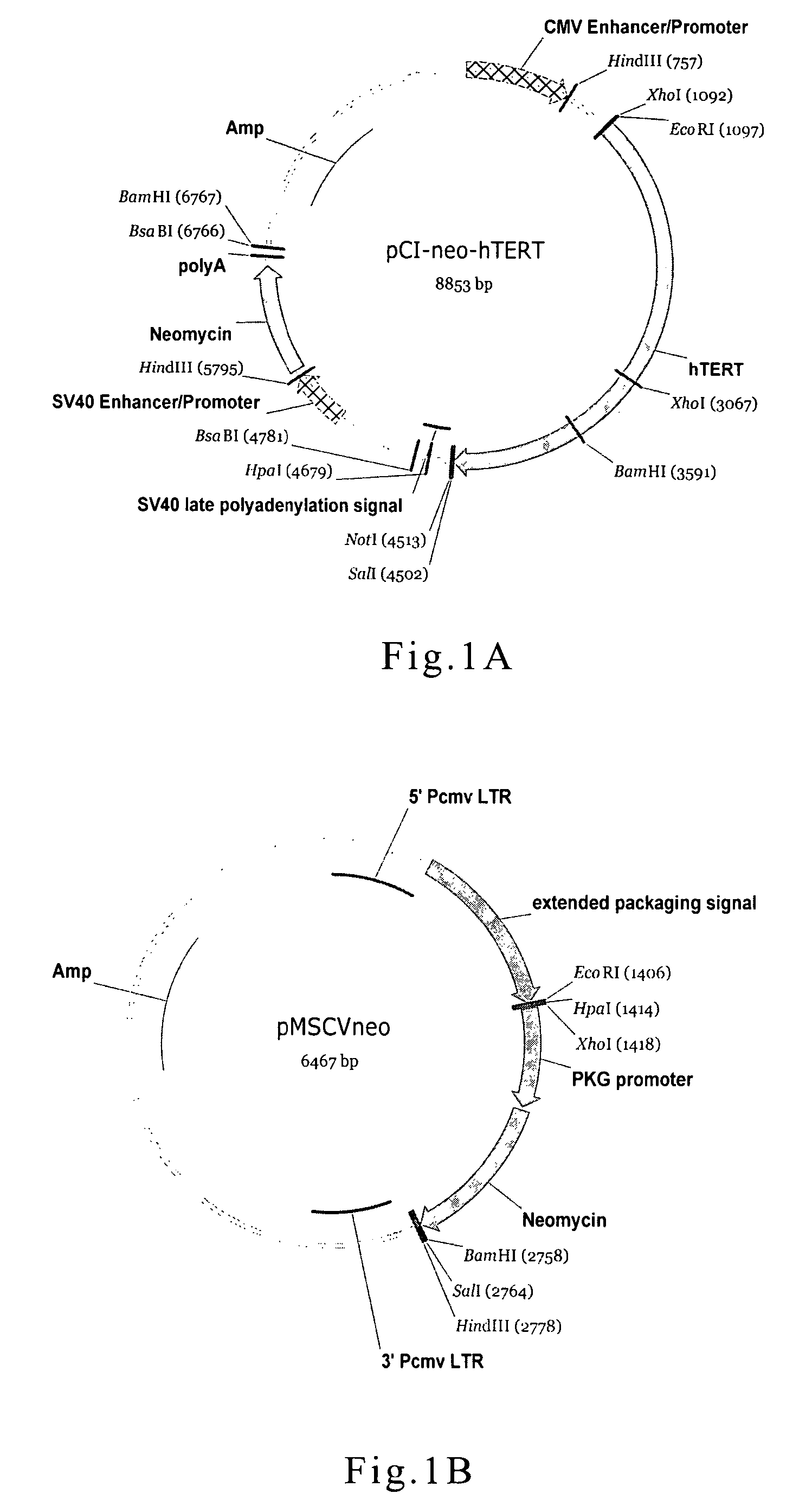
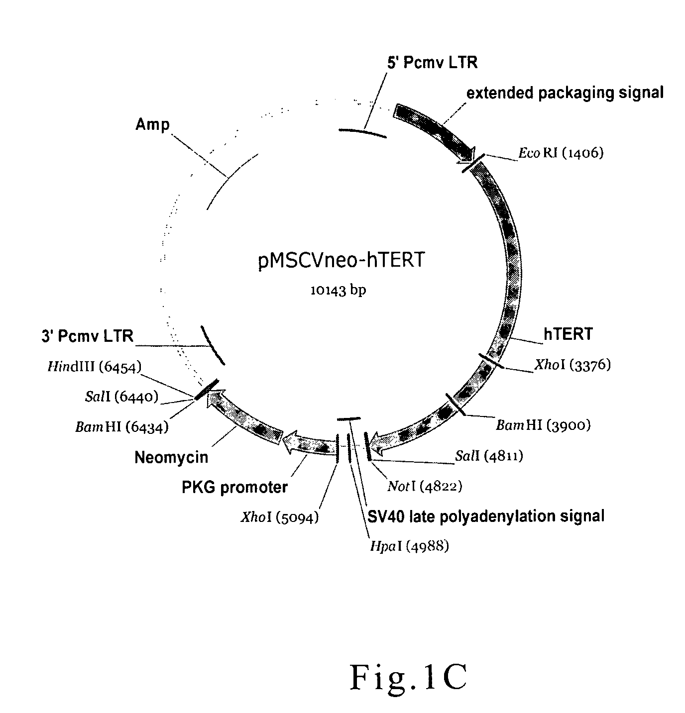
[0048]In the present example, one of the cell lines used in the cell model was human HSC, which was transfected with virus particles carrying immortalizing vector. The plasmid pC1-neo hTERT (FIG. 1A, Promega Corporation) was restriction digested with EcoR I and Sat I, and pMSCV-neo (FIG. 1B, Clontech Laboratories, Inc.) plasmid was also digested with EcoR I and Xho I. The digested fragments were separated by electrophoresis, and desired fragments were purified with QIA quick Gel Extraction Kit (QIAGEN). Purified fragments were ligated via T4 DNA ligase, and the retroviral vector of pMSCV-neo hTERT was thus obtained (FIG. 1C).

[0049]The retroviral vector of pMSCV-neo hTERT constructed above was transfected into packing cells—RetroPack™pT67 or AmphoPack™-293 by Lipofectamine Reagents (Invitrogen Corporation). Culture medium containing retrovirus particles were collected and used to inf...

example 2

Construction of Retroviral Vector Containing Albumin Promoter

[0051]In the present example, hepatocyte cell line used in the cell model was Huh-7 cells transfected with retroviral vector containing albumin promoter. The transfected cells in the present invention were selected by hygromycin. Besides the hygromycin resistant gene, the retroviral vector also contained a liver cell specific albumin promoter and an enhanced green fluorescence protein (EGFP) reporter gene.

[0052]To clone albumin promoter, the sequence of accession number 00178343 in GenBank was used to design primers hSA-3(SEQ ID NO.1) and hSA-5(SEQ ID NO.2). RT-PCR was performed with primers hSA-3 and hSA-5 to amplify albumin promoter fragment from human cDNA, the sequence of the amplified fragment is shown in SEQ ID NO.3.

[0053]The gene fragment of the enhanced green fluorescence protein (EGFP) was amplified with two primers of EGFP-1 (SEQ ID NO. 4) and EGFP-3 (SEQ ID NO. 5) from plasmid pEGFP-1 (FIG. 3A, Clontech Laborato...

example 3

Construction of Retroviral Vector Carrying α-I-antitrypsin Promoter

[0055]In the present example, the cells used for the cell model can be Huh-7 cells transfected by a retrovirus particles carrying α-I-antitrypsin promoter. Besides liver cell specific promoter—α-I-antitrypsin promoter, the retroviral vector also encodes hygromicin resistant gene and EGFP gene.

[0056]The sequence of accession number 00177830 in GenBank containing liver specific α-I-antitrypsin promoter, which was used to design primers AAT-1 (SEQ ID NO.8) and AAT-2 (SEQ ID NO.9). RT-PCR was performed with primers AAT-1 and AAT-2 to amplify α-I-antitrypsin promoter fragment from human cDNA, and the sequence of the amplified fragment is shown in SEQ ID NO.10.

[0057]As described in example 1, the EGFP gene was amplified with two primers—EGFP-1 (SEQ ID NO: 4) and EGFP-3 (SEQ ID NO: 5) from plasmid pEGFP-1 (FIG. 3A, Clontech Laboratories, Inc.). The hygromicin resistant gene was amplified from plasmid pMSCV-hyg (FIG. 3B, Clo...


PUM

PropertyMeasurementUnit
emission wavelengthaaaaaaaaaa
excitation wavelengthaaaaaaaaaa
emission wavelengthaaaaaaaaaa

Abstract

The present invention provides a cell-culturing model and a method for screening compounds which can be applied in treating or preventing hepatic cirrhosis. The cell culturing model comprises a population of hepatocytes and hepatic stellate cells (HSCs) derived from co-culturing, and at least one population of the cells comprises a nucleotide sequence fragment of a reporter gene and a cell specific regulatory sequence. The cell-culturing model of the present invention can be applied in high throughput screening for effective compounds of medication, and also in understanding the functional mechanism of the effective compounds.

Description

BACKGROUND OF THE INVENTION[0001]1. Field of the Invention[0002]The present invention relates to a cell culturing model and a method for drug screening, more particularly, to a cell culturing model and a method using modified liver cells and hepatic stellate cells in high throughput drug screening for hepatic cirrhosis treatment.[0003]2. Description of Related Art[0004]Hepatic cirrhosis is often resulted from the damage to hepatocytes by alcohol and drugs, or virus infection. Damaged hepatocytes release certain signals to activate hepatic stellate cells (HSCs) in liver tissue into myofibroblasts (MFs). MFs are capable of secreting large varieties of extracellular matrix (ECM), including collagen, fibronectin, laminin and proteoglycan. The accumulation of excess extracellular matrix causes liver fibrosis. In the meantime, accumulated extracellular matrix further obstructs hepatocytes from the supply of oxygen and nutrients, and therefore leads to necrosis. To sum up, hepatic cirrhosi...

Claims


Application Information

Patent Timeline
Patent Type & Authority Patents(United States)
IPC IPC(8): C12Q1/68C12N5/071C12N5/00C12Q1/66
CPCG01N33/5023G01N33/5067G01N2800/085
Inventor LI, WEN-TYNGGAU, RUNG-JIUNWENG, YU-SHIHHSIEH, CHENG-TALI, PEI-SHAN
Owner IND TECH RES INST
Learn More