Micromachined bilayer unit for filtration of small molecules

a micro-machined bilayer unit and filtration technology, applied in the field of organ transplantation and reconstructive surgery, can solve the problems of distressing dialysis patients, high invasiveness, and limited effectiveness of dialysis patients

Inactive Publication Date: 2010-08-17
THE GENERAL HOSPITAL CORP +2
View PDF88 Cites 69 Cited by
  • Summary
  • Abstract
  • Description
  • Claims
  • Application Information

AI Technical Summary

Benefits of technology

[0018]In yet another embodiment, the first and / or second molds can optionally include flow control patterns, such as interconnected microchannels cut into the mold, or disposed on a surface thereof. As used herein, the term “flow control pattern” refers to any feature capable of affecting flow, e.g., limiting or directing fluid flow, ranging from two-dimensional surface patterns to three-dimensional features such as posts, walls, channels or other raised structures. The flow control pattern can enhance diffusion and convection, among other things. The patterns in the first mold can be identical or non-identical to the pattern in the second mold. The pattern in the upper surface of the molds can be identical or non-identical to the pattern in the lower surface.
[0026]The surfaces of the molds are biocompatible can include a coating on the microchannels that promotes cell adhesion to the apparatus. The surfaces are preferably hemocompatible, or treated to promote hemocompatibility by, for example, the application of a hemocompatible film thereto or endothelialization of the surfaces. Blood clot-preventing compounds such as heparin can also be used to further facilitate the uninhibited flow of blood and long-term operation of the device, among other things.
[0032]In the aforementioned method, the step of allowing the polymeric liquid in contact with the mold and membrane to set can further include elevating the surrounding temperature.

Problems solved by technology

These treatments, while effective at sustaining life for ESRD (End-Stage Renal Disease) patients, are highly invasive, and are limited in effectiveness due to the non-physiological concentration profile of waste and impurities in the bloodstream.
These drastic excursions in concentration lead to complications and distress in dialysis patients, and the psychological impact of dialysis is associated with a sharp increase in suicide rates among the patient population.
Most significantly, the long-term prognosis for ESRD patients on dialysis is poor, with 5-year survival rates of less than 20%.
The cost of these treatments is staggering, totaling approximately $12 Billion per year for the roughly 300,000 ESRD patients, or $40,000 per year per patient.
These sources include cadaveric organs, which are in extremely limited supply, and therefore the number of patients on the waiting list for a vital organ is approaching 100,000 in the United States.
Organ rejection by the recipient's immune system represents a huge challenge for the field of transplant medicine, because it severely limits the potential donor pool.
Even when the donor is a match, recipients are consigned to a lifetime of immunosuppressive drugs, which are extremely expensive and are associated with a host of severe side effects.
Often involving family members, these transplants are typically safe for the donor but have led to well-documented cases in which previously healthy donors suffered lethal complications following transplant surgery.
The principal disadvantages of the two general approaches described above relate to the insufficient replacement of physiological organ function without serious limitations or complications.
In the case of renal assist devices, specifically, the invasive, complex and discontinuous nature of the treatments limit their therapeutic value, because they do not provide patients with benefits concomitant with a healthy pair of kidneys.
These disadvantages can be understood as being associated with limitations in the technology which insufficiently reproduce organ function, and limitations associated with cost, complexity and accessibility.
The latter set of challenges relates to the fact that dialysis treatments are costly and labor-intensive, require frequent visits to the clinic and large, complex machines, and are not continuous because of the need for centralized dialysis clinics often distant from their patients.

Method used


Image

  • Micromachined bilayer unit for filtration of small molecules
  • Micromachined bilayer unit for filtration of small molecules
  • Micromachined bilayer unit for filtration of small molecules

Examples


examples and specific embodiments

[0115]Results obtained on single bilayer devices tested for ultrafiltration of urea and creatinine are described in the following discussion. Single bilayer devices were constructed using some or all of the techniques described in the above fabrication section.

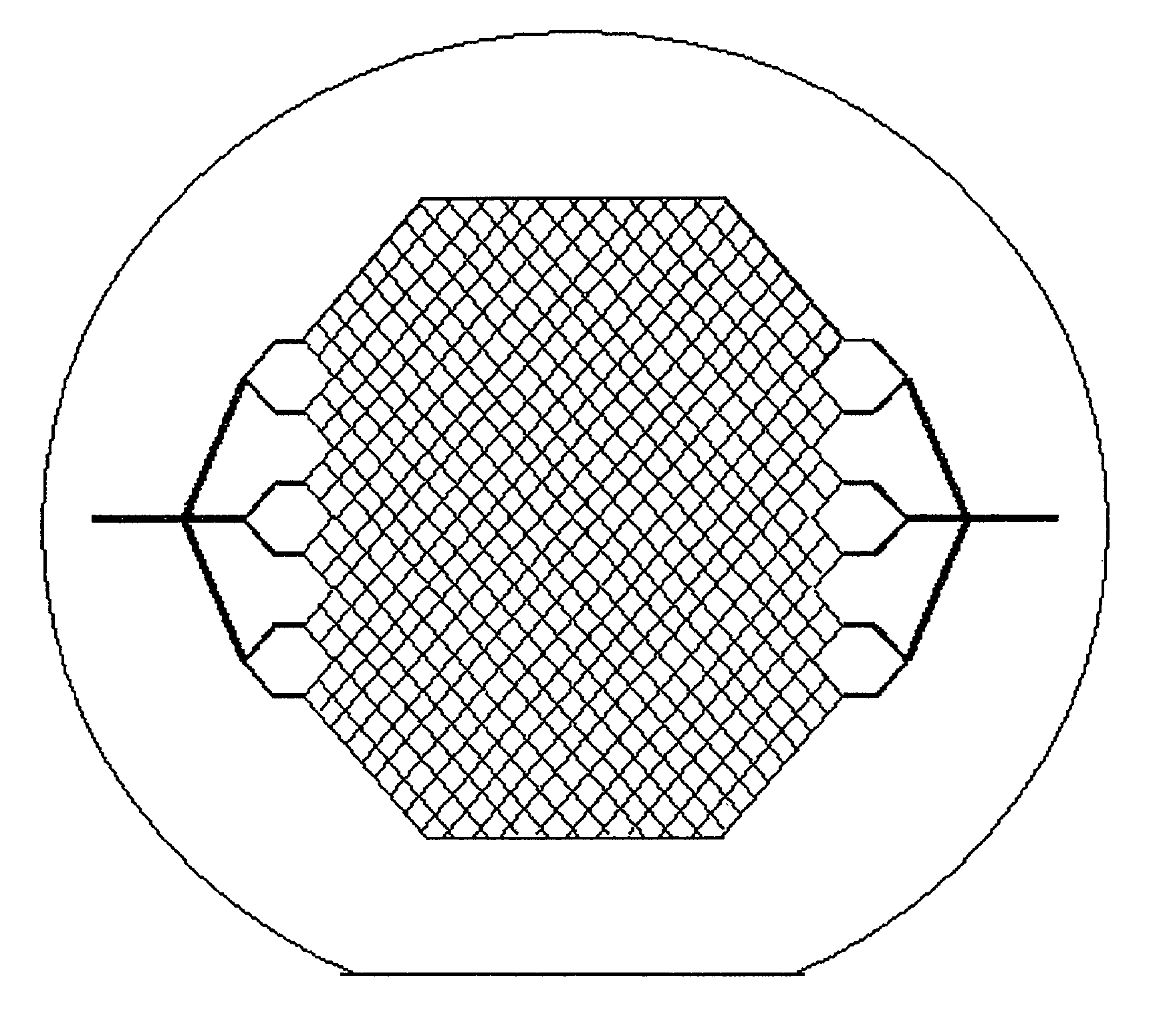
[0116]A device 110 of the present invention is shown schematically in FIG. 4. Device 110 includes a first mold layer 118a and a second layer 118b separated by a microporoous membrane 122. In one embodiment, the first mold layer 118a can comprise a microvascular network, including microchannels that direct flow through chambers and conduits, and the second mold layer 118b can comprise a parenchymal layer having cells. The term “microvascular network,” as used herein, refers to fluidic network modeled after a physiologic vasculature, such as a capillary network. The microvascular network either may or may not consist of an actual endothelium. In another embodiment, the mold layers are acellular (e.g., no cells are cultured withi...


PUM

PropertyMeasurementUnit
heightaaaaaaaaaa
widthaaaaaaaaaa
widthaaaaaaaaaa

Abstract

Methods and materials for making an apparatus which duplicates the functionality of a physiological system id provided.

Description

CROSS REFERENCE TO RELATED APPLICATION(S)[0001]Reference is made to U.S. application Ser. No. 10 / 038,891, filed Jan. 2, 2002, which claims priority to U.S. Appln. Ser. No. 60 / 259,283, filed Jan. 2, 2001. Reference is made to U.S. application Ser. No. 09 / 560,480, filed Apr. 28, 2000, which claims priority to U.S. Appln. Ser. No. 60 / 131,930 filed Apr. 30, 1999, and U.S. Appln. Ser. No. 60 / 165,329, filed Nov. 12, 1999. The contents of each of these applications are incorporated by reference herein in their entireties.STATEMENT REGARDING POTENTIAL GOVERNMENT INTEREST[0002]The United States government has certain rights in this invention by virtue of grant number DAMD-17-02-2-0006 from the Department of the Army.INCORPORATION BY REFERENCE[0003]Each of the foregoing applications-and patents and articles, and each document cited or referenced in each of the foregoing applications and patents and articles, including during the prosecution of each of the foregoing applications and patents (“...

Claims


Application Information

Patent Timeline
Patent Type & Authority Patents(United States)
IPC IPC(8): C12M3/06C12M1/34C12N5/08
CPCA61M1/1678Y10T436/25375Y10T436/255
Inventor BORENSTEIN, JEFFREY T.WEINBERG, ELIORRICK, BRIAN K.PRITCHARD, ELEANOR M.BARNARD, EDWARD J.KREBS, NICHOLAS J.MARENTIS, THEODOREVACANTI, JOSEPH P.KAAZEMPUR-MOFRAD, MOHAMMAD REZA
Owner THE GENERAL HOSPITAL CORP
Why Eureka
Try Eureka