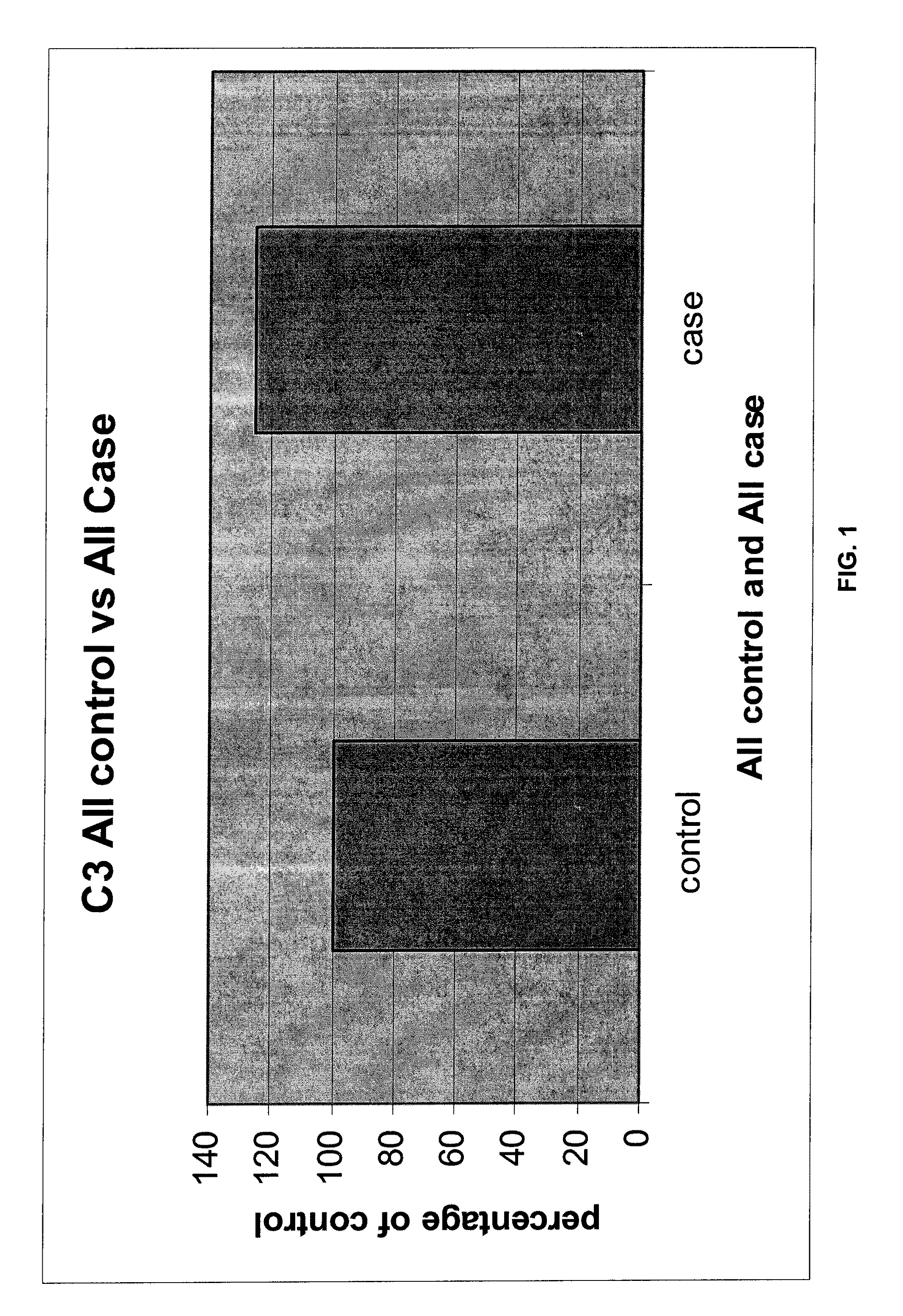
Serum biomarkers for early detection of acute cellular rejection
a cellular rejection and serum biomarker technology, applied in the field of acute cellular rejection diagnosis and prevention, can solve the problem of difficult identification of the effects of hcv and acr
- Summary
- Abstract
- Description
- Claims
- Application Information
AI Technical Summary
Benefits of technology
Problems solved by technology
Method used
Image
Examples
example 1
Preventing ACR by Monitoring Immunosuppression
[0074]A subject who received a solid organ (e.g. heart, liver or kidney) allograft (transplant) four weeks ago is usually maintained on a cocktail of immunosuppressive agents. The immunosuppressive agents might include a calcineurin inhibitor, a corticosteroid and an antiproliferative agent. The purpose of prescribing these agents is to prevent allograft injury through rejection.
[0075]All immunosuppressive agents have important side effects and toxicities. Thus, a careful balance between effective dosing and toxicity must be struck for each subject receiving immunosuppressive therapy. Currently, physicians are guided by either drug levels (e.g. for tacrolimus or mycophenolate mofetil) or simple dosing schedules (e.g. for corticosteroids). Knowing the state of immunological activation, as indicated by the differential abundance of one or more of the proteins associated with acute cellular rejection, enables a physician to adjust dosing of...
example 2
Monitoring Immune Activation
[0078]Because ACR can cause allograft injury that is indistinguishable from other causes of allograft injury, such as infection or loss of blood supply, measuring serum proteins differentially associated with ACR provides important diagnostic information. For example, a liver transplant recipient might present two months post transplantation with elevated bilirubin and aninotransferase levels. The subject is known to have hepatitis C (HCV) infection. Biopsy findings associated with HCV and ACR can overlap substantially. Levels of ubiquitin conjugating enzyme E2 and beta-2-glycoprotein 1 precursor, measured by multiplex enzyme-linked immunosorbance assay (ELISA) are found to be compatible with ACR when compared to a standardized upper limit of normal for subjects who do not develop ACR. With this knowledge, treatment might focus on increasing immunosuppression for this subject, thereby preventing the subject from developing ACR. Treating HCV in this settin...
example 3
Diagnosing Acute or Incipient Acute Cellular Rejection
[0079]ACR occurs with varying frequencies depending on the organ transplanted. The frequency also varies with host factors, such as age, nutritional status and cause of transplanted organ failure. The development of ACR can adversely affect any transplanted organ, with consequences that can include death or graft loss.
[0080]Currently, diagnosing ACR is based on a composite of clinical picture (e.g. rising creatinine for kidney transplant recipients, or rising liver biochemistries for liver transplant recipients) and histology. Histological examination of an allograft necessitates biopsy of the affected organ. Biopsies are expensive, invasive and potentially dangerous procedures. In contrast, measuring serum proteins according to the present invention that are specifically differentially associated with ACR could negate the need for organ biopsies to diagnose rejection (or to determine response to anti-rejection treatment).
[0081]F...
PUM
| Property | Measurement | Unit |
|---|---|---|
| time | aaaaa | aaaaa |
| molecular weight | aaaaa | aaaaa |
| crystal structure | aaaaa | aaaaa |
Abstract
Description
Claims
Application Information
 - R&D
- Intellectual Property
- Life Sciences
- Materials
- Tech Scout
- Unparalleled Data Quality
- Higher Quality Content
- 60% Fewer Hallucinations
Browse by: Latest US Patents, China's latest patents, Technical Efficacy Thesaurus, Application Domain, Technology Topic, Popular Technical Reports.
© 2025 PatSnap. All rights reserved.Legal|Privacy policy|Modern Slavery Act Transparency Statement|Sitemap|About US| Contact US: help@patsnap.com

