Hyperspectral imaging in diabetes and peripheral vascular disease

a multi-spectral imaging and diabetes technology, applied in the field of hyperspectral and multi-spectral imaging of medical tissues, can solve the problems of poor drugcologic treatment of pads, unsatisfactory relying on medial history and physical examination alone, and about 10% of people, so as to evaluate the potential for wound healing and overcome the problems and disadvantages

Active Publication Date: 2013-02-12
HYPERMED IMAGING
View PDF50 Cites 39 Cited by
  • Summary
  • Abstract
  • Description
  • Claims
  • Application Information

AI Technical Summary

Problems solved by technology

Problems with the foot that affect the ambulatory nature of the patient are not only important from the standpoint of physical risk, but also convey an emotional risk as well, as these problems disrupt the fundamental independence of the patient by limiting his or her ability to walk.
Although pharmacologic treatments for PAD have traditionally been poor, 2.1 million nevertheless receive pharmacologic treatment for the symptoms of PAD, and current diagnostic tests are not considered to be very sensitive indicators of disease progression or response to therapy.
One difficulty in diagnosing PAD is that in the general population, only about 10% of persons with PAD experience classic symptoms of intermittent claudication.
Relying on medial history and physical examination alone is unsatisfactory.
As in the case of diabetic foot disease, current technologies have fallen short.
Another problem face by physicians is disease of the peripheral veins.
Venous occlusive disease due to incompetent valves in veins designed to prevent backflow and deep vein thrombosis, results in venous congestion and eventually stasis ulcers.
This, in turn, can lead to increased pressure on the dermis, resulting in tissue ischemia and eventual death, and ultimately result in an ulcer.4 There are a number of factors that weigh heavily in the process of ulcerations5—affecting different aspects of the foot—that lead to a combination of effects that greatly increase the risk of ulceration:6 Neuropathy—Results in a loss of protective sensation in the foot, exposing patients to undue, sudden or repetitive stress.
Can cause a lack of awareness of damage to the foot as it be occurs and physical defects and deformities7 which lead to even greater physical stresses on the foot.
It can also lead to increased risk of cracking and the development of fissures in calluses, creating a potential entry for bacteria and increased risk of infection.8 Microcirculatory Changes—Often seen in association with hyperglycemic damage9 Functional abnormalities occur at several levels, including hyaline basement membrane thickening and capillary leakage.
On a histologic level, it is well known that diabetes causes a thickening of the endothelial basement membrane which in turn may lead to impaired endothelial cell function.Musculoskeletal Abnormalities—Include altered foot mechanics, limited joint mobility, and bony deformities, and can lead to harmful changes in biomechanics and gait.
This increases pressures associated with various regions of the foot.
Alteration or atrophy of fat pads from increased pressure can lead to skin loss or callus, both of which increase the risk of ulceration by two orders of magnitude.Peripheral Vascular Disease—Caused by atherosclerotic obstruction of large vessels resulting in arterial insufficiency10 is common in the elderly populations and is yet more common and severe in diabetics.11 Diabetics may develop atherosclerotic disease of large-sized and medium-sized arteries, however, significant atherosclerotic disease of the infrapopliteal segments is particularly common.
Foot pathology is major source of morbidity among diabetics and is a leading cause of hospitalization.
None of these methods are discriminatory for feet at risk, and none of them provide any information about the spatial variability across the foot.
Currently there is no method to accurately assess the predisposition to serious foot complications, to define the real extent of disease or to track the efficacy of therapeutics over time.
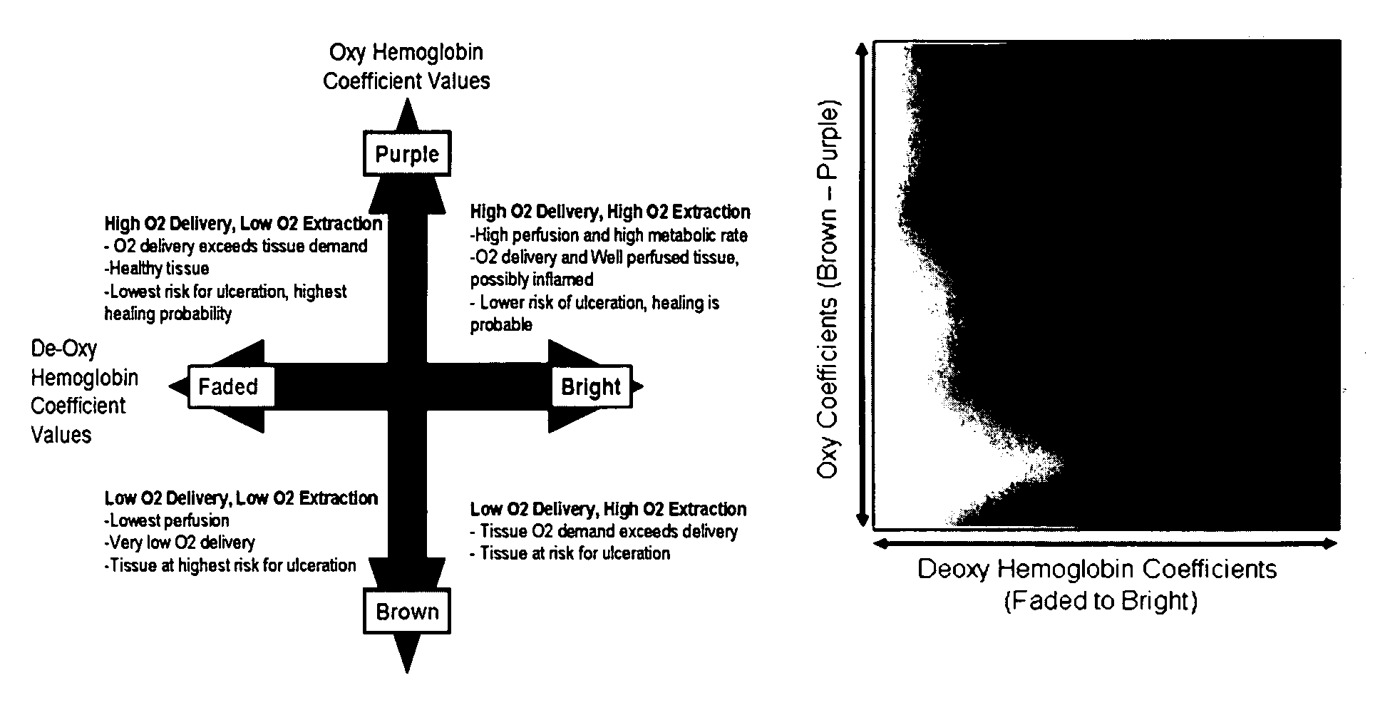
One of the most common spectroscopic applications is in pulse oximetry, which utilize the different oxyhemoglobin (oxyHb) and deoxyhemoglobin (deoxyHb) absorption bands to estimate arterial hemoglobin oxygen saturation.28 One of the drawbacks of these systems is that they provide no information about the spatial distribution or heterogeneity of the data.
In addition, these systems report the ratio of oxyHb and deoxyHb together losing diagnostic information that can be garnered by evaluating the state of the individual components.
TcPO2 measurements appeared cumbersome, lengthy (˜20-30 minutes), highly operator dependant, and carried data only from skin directly under the probe (with little ability to distinguish the spatial characteristics of the ischemic area).
While TcPO2 has been shown to carry statistically significant information in terms of quantifying tissue at risk for ulceration,48 TcPO2 was not encouraging as a useful clinical device.Non-imaging techniques—Techniques such as near-infrared absorption spectroscopy (NIRS) or TcPO2, rely on measurements at a single point in tissue which may not accurately reflect overall tissue condition or provide anatomically relevant data, and probe placement on the skin can alter blood flow and cannot deliver accurate information in the area of an ulcer or directly surrounding it.
Because MHSI is truly remote sensing, data are acquired at a distance, eliminating probe placement errors and allowing the investigation of the wound itself, which some techniques can not accomplish due to infection risk.
Hence, the discrimination is not markedly improved by adding iontophoresis results to refine prediction as is required for Laser Doppler to do so.
Laser Doppler data has poor spatial resolution and is frequently reported as a single mean numerical value across the region of interest.

Method used


Image

  • Hyperspectral imaging in diabetes and peripheral vascular disease
  • Hyperspectral imaging in diabetes and peripheral vascular disease
  • Hyperspectral imaging in diabetes and peripheral vascular disease

Examples


Embodiment Construction

[0062]The major clinical advantage of hyperspectral imaging is the delivery of metabolic information derived from the tissue's spectral properties in an easily interpretable image format with high spatial resolution. This 2-D information allows gradients in biomarker levels to be assessed spatially. Multiple images taken over time allow the gradient to be measured temporally. This adds new dimensions to the assessment of ulceration risk and tissue healing in that it will allow the physician to target therapy and care to specific at risk areas much earlier than previously possible. The reporting of biomarkers such as oxyHb and deoxyHb levels in tissue individually and in an image format where spatial distributions can be assessed has not been done before. Typically the two numbers are combined in a ratio and reported as percent hemoglobin oxygen saturation (O2Sat). MHSI has the clear potential to be developed into a cost effective, easy to use, turn-key camera-based metabolic sensor ...


PUM


Abstract

The invention is directed to methods and systems of hyperspectral and multispectral imaging of medical tissues. In particular, the invention is directed to new devices, tools and processes for the detection and evaluation of diseases and disorders such as, but not limited to diabetes and peripheral vascular disease, that incorporate hyperspectral or multispectral imaging.

Description

REFERENCE TO RELATED APPLICATIONS[0001]The present application claims priority to U.S. Provisional Patent Application Ser. No. 60 / 667,677 entitled Hyperspectral Imaging in Diabetes, filed Apr. 4, 2005, and U.S. Provisional Patent Application Ser. No. 60 / 785,977 entitled Hyperspectral Imaging of Angiogenesis, filed Mar. 27, 2006 which is hereby incorporated by reference.BACKGROUND[0002]1. Field of the Invention[0003]The invention is directed to methods and systems of hyperspectral and multispectral imaging of medical tissues. In particular, the invention is directed to new devices, tools and processes for the detection and evaluation of diseases and disorders such as diabetes and peripheral vascular disease that incorporate hypespectral / multispeatral imaging.[0004]2. Background of the Invention[0005]Diabetes afflicts an estimated 194 million people worldwide, affecting 7.90% of Americans (over 21 million people) and 7.8% of Europeans. Between 85% and 95% of all diabetics suffer from ...

Claims


Application Information

Patent Timeline
Patent Type & Authority Patents(United States)
IPC IPC(8): A61B6/00
CPCA61B5/0059G06T7/0012A61B5/14552A61B5/412A61B5/444A61B5/445A61B5/7275G01J3/02G01J3/0224G01J3/0272G01J3/0283G01J3/10G01J3/12G01J3/2823A61B5/145A61B5/743A61B5/14551G16H30/40G16H50/20
Inventor FREEMAN, JENNY E.PANASYUK, SVETLANA V.HOPMEIER, MICHAEL J.SCHOMACKER, KEVINBRAND, DEREK
Owner HYPERMED IMAGING
Why Eureka
Try Eureka