
Optical assembly and method for determining analyte concentration

an analyte concentration and optical assembly technology, applied in the field of optical assembly and method for determining the concentration of analyte in body fluid, can solve the problems of poor patient acceptance, inability to read during the night or when the patient is otherwise occupied, and impose a considerable burden on the diabetic patien

Inactive Publication Date: 2015-03-03
MELYS DIAGNOSTICS
View PDF20 Cites 0 Cited by
  • Summary
  • Abstract
  • Description
  • Claims
  • Application Information

AI Technical Summary

Benefits of technology

The patent text describes a method for improving the penetration of light into tissue samples for absorption by the analyte. This is achieved by using a second element with a larger refractive index than the first element and the tissue sample, which causes the light to incident at an angle less than the critical angle for total internal reflection. Another embodiment of the invention is an interferometer with two reflecting surfaces, where the front surface of each element has an anti-reflection coating to minimize light reflection. Both methods ensure that the light penetrates beyond the stratum corneum to the required depth for absorption by the analyte.

Problems solved by technology

The main disadvantages of self blood glucose monitoring systems are poor patient acceptance because the technique is painful, only intermittent assessment of diabetic control is possible and readings during the night or when the patient is otherwise occupied are not possible.
This increase in the frequency of blood glucose testing imposes a considerable burden on the diabetic patient, both in financial terms and in terms of pain and discomfort.
Disadvantages of the use of NIR spectroscopy are that the measurement suffers from interference by other optical absorbers in the tissue and is also dependent on blood flow in the skin.
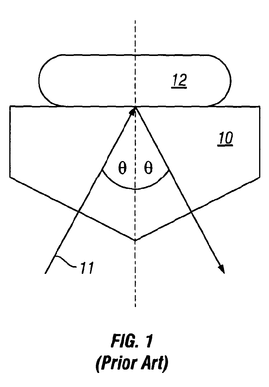
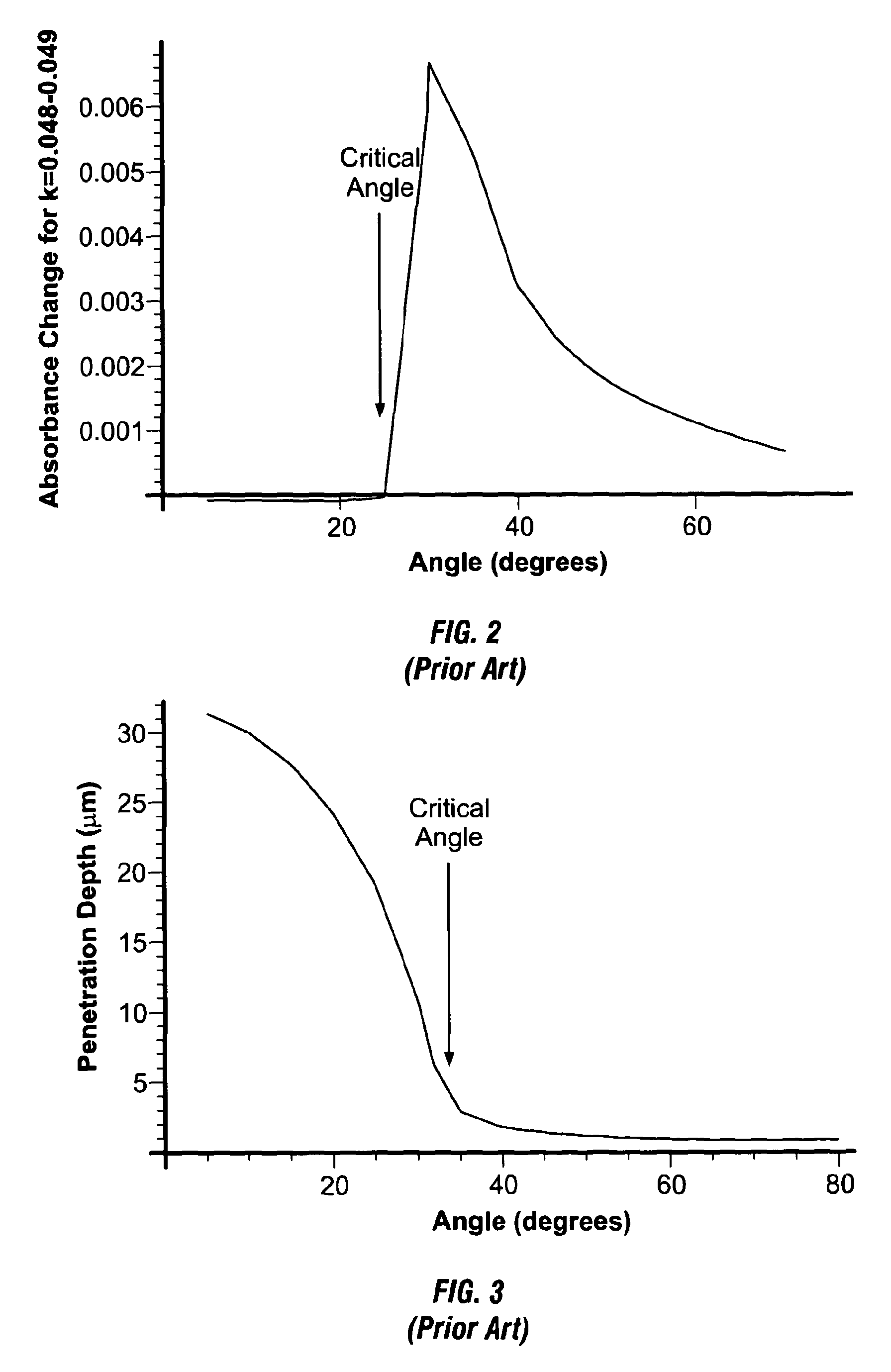
However, at the critical angle the value of δ is typically only 3 μm so that the effective penetration depth for most subjects is insufficient for making a measurement of analyte concentration.
It is for these reasons that conventional ATR cannot measure glucose and other analyte concentration in the majority of subjects non-invasively.

Method used


Image

  • Optical assembly and method for determining analyte concentration
  • Optical assembly and method for determining analyte concentration
  • Optical assembly and method for determining analyte concentration

Examples


first embodiment

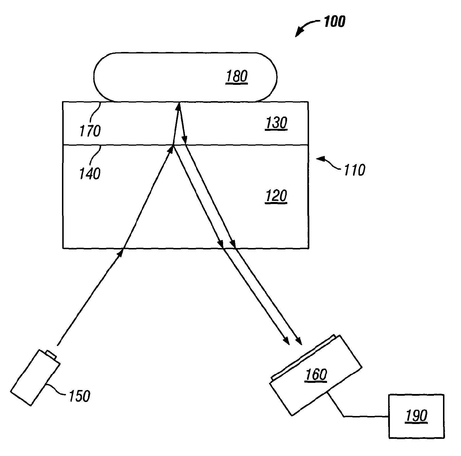
[0052]Referring to FIG. 4 of the drawings, there is illustrated an optical assembly 100a according to the present invention comprising an optical arrangement 110a, for the non-invasive determination of the concentration of an analyte, such as glucose, in a tissue sample, such as body fluid.

[0053]The optical arrangement 110a illustrated comprises a substantially rectangular cuboid shape however, it is to be appreciated that other shapes may also be used. The arrangement comprises a first, relatively thick layer 120 of calcium fluoride, for example and a second, relatively thin layer 130 of parylene for example, disposed upon the first layer 120 and coupled thereto to form an interface 140 between the first and second layers 120, 130. The first and second layers 120, 130 are substantially transmissive to light in the infra-red region of the spectrum, namely in the wavelength range 1-10 μm, such that light in this wavelength range can pass through the first and second layers 120, 130.

[...

second embodiment

[0064]Referring to FIG. 7 of the drawings, there is illustrated an optical assembly 200 according to the present invention for the non-invasive determination of the concentration of an analyte, such as glucose, in a tissue sample. The assembly 200 comprises an optical arrangement 210 and a broadband light source 150, such as a broadband laser which is arranged to launch laser light having a wavelength characteristic of infra-red wavelengths. The optical arrangement 210 comprises an interferometer comprising a beam splitter 220 which is arranged to partially reflect and partially transmit incident laser light toward a first and second reflector element 230, 240, respectively.

[0065]The reflector elements 230, 240 may be formed of zinc selenide and comprise substantially planar element. The elements 230, 240 are orientated to reflect the light incident thereon back toward the beam splitter 220, substantially along the same path as the incident light. In this respect, the elements 230, ...


PUM

PropertyMeasurementUnit
concentrationaaaaaaaaaa
thicknessaaaaaaaaaa
wavelength rangeaaaaaaaaaa

Abstract

An optical assembly and method for the non-invasive determination of a concentration of an analyte in a tissue sample is disclosed. The assembly comprises an optical arrangement comprising a first optical interface for reflecting light incident thereon and a second optical interface for reflecting light incident thereon. The second interface comprises an interface between an optical element of the arrangement and the tissue sample under investigation. The light reflected from the first and second interfaces is arranged to combine to generate an interference pattern characteristic of a difference in phase between light reflected from the first interface with the light reflected from the second interface.

Description

CROSS-REFERENCE TO RELATED APPLICATIONS[0001]This application is a national stage application of PCT / GB2011 / 051246 filed on Jul. 1, 2011 and claims priority from GB 1011459.3, filed on Jul. 7, 2010 and from GB 1105045.7, filed on Mar. 25, 2011, which are hereby incorporated by reference in their entireties.BACKGROUND[0002]1. Technical Field[0003]The present invention relates to an optical assembly and method for determining the concentration of an analyte in body fluid and particularly, but not exclusively, to an optical assembly and method for determining the concentration of glucose in body fluid.[0004]2. Related Art[0005]Diabetes mellitus, which is commonly known as diabetes, is the name for a group of chronic diseases that affect the way the body uses food to make the energy necessary for life. Diabetes is primarily a disruption of carbohydrate metabolism with the result that the blood glucose level of a person may vary considerably, from 40 mM (720 mg / dl) to as low as 1 mM (18 ...

Claims


Application Information

Patent Timeline
Patent Type & Authority Patents(United States)
IPC IPC(8): G01N21/55A61B5/145A61B5/1455G01N21/35G01N21/552
CPCA61B5/14532A61B5/1455G01N21/55G01N21/552G01N21/35
Inventor ENDERBY, JOHNPARKER, DAWOOD
Owner MELYS DIAGNOSTICS
Learn More