Method for decellularization of tissue grafts

a tissue graft and decellularization technology, applied in the field of tissue graft decellularization, can solve the problems of curtail nerve regeneration, routinely involved rigorous decellularization techniques, loss of clean ends for direct repair or gap, etc., to reduce the cd4+ t-cell mediated immune response, promote graft implantation, and reduce the effect of recipient immune respons

Active Publication Date: 2017-02-21
UNIV OF FLORIDA RES FOUNDATION INC
View PDF7 Cites 8 Cited by
  • Summary
  • Abstract
  • Description
  • Claims
  • Application Information

AI Technical Summary

Benefits of technology

[0011]The tissue grafts of the present invention provide a natural replacement tissue or graft that can be obtained via simple preparation steps. The methods of the invention specifically disrupt immunogenic cellular components without significantly altering the natural extracellular structure. Advantageously, the tissue retains biological properties for promoting cellular regeneration through the graft. The native extracellular matrix (ECM) structure is preserved, specifically, in the case of nerve grafts, the basal laminae and endoneurium / endothelial layer retain their natural and generally original structure.
[0013]In one embodiment, the present invention provides a method for preparing tissue replacement that includes the steps of: washing the replacement tissue, in the absence of any ionic detergents (including any anionic detergent), in a solution comprising at least one amphoteric detergent and then washing the replacement tissue in serial solutions of a buffered salt to remove excess amphoteric detergent. This process is simpler and less expensive than processes of the current art and results in a replacement tissue with better handling properties and with better regeneration-promoting properties.

Problems solved by technology

Biomedical engineering faces many challenges in the development of tissue that can be used to facilitate healing.
One particularly challenging embodiment of tissue engineering is the production of nerve grafts that can be used to help regeneration of severed nerves.
In part because of this process, axonal and myelin debris have long been believed to have growth-inhibitory effects that curtail nerve regeneration and may be a mechanical barrier to axonal growth.
Consequently, processing methods used for nerve grafts have routinely involved rigorous decellularization techniques, even at the expense of disrupting extracellular matrix (ECM) structures and nerve graft integrity that support the regenerative process.
Nerve damage often results in the loss of clean ends for direct repair or a gap created by nerve tissue damage.

Method used


Image

  • Method for decellularization of tissue grafts
  • Method for decellularization of tissue grafts
  • Method for decellularization of tissue grafts

Examples


example 1

Decellularization Process

[0083]All procedures should employ aseptic techniques and sterile solutions. Following are the steps of one embodiment of the decellularization process of the subject inventon.

1. Resect nerve from a donor and store frozen.

2. Thaw the nerve at room temperature. Rinse with chilled water. Submerge in a large volume of chilled water. Gently agitate at 4° C. for 6 hrs (or longer).

[0084]3. Submerge the nerves in a large volume of 125 mM (or more concentrated) Sulfobetaine-10 in Extraction Buffer (10 mM Na phosphate buffer, pH 7.2+150 mM NaCl) or any physiologic saline. Gently agitate at 25° C. (near room temperature) for 15 hrs (or longer).

4. Replace with a large volume of fresh 125 mM (or more concentrated) Sulfobetaine-10 in Extraction Buffer or any physiologic saline. Agitate for 6 hrs (or longer) at 25° C.

5. Wash with a large volume of Extraction Buffer or any physiologic saline. Agitate at 25° C. for 60 min (or longer).

6. Wash with a large volume of Rinse Buf...

example 2

Comparison of Decellularization Method with Rabbit Nerves

[0085]Grafts prepared in accordance with the subject invention as described above are referred to as DC3 grafts. DC3 grafts were compared to normal (unprocessed) rabbit nerve and rabbit grafts processed by the protocol described by Hudson et al., (Tissue Engineering 10:1346-1358, 2004) and U.S. Pat. No. 7,402,319; referred to herein as DC2 grafts.

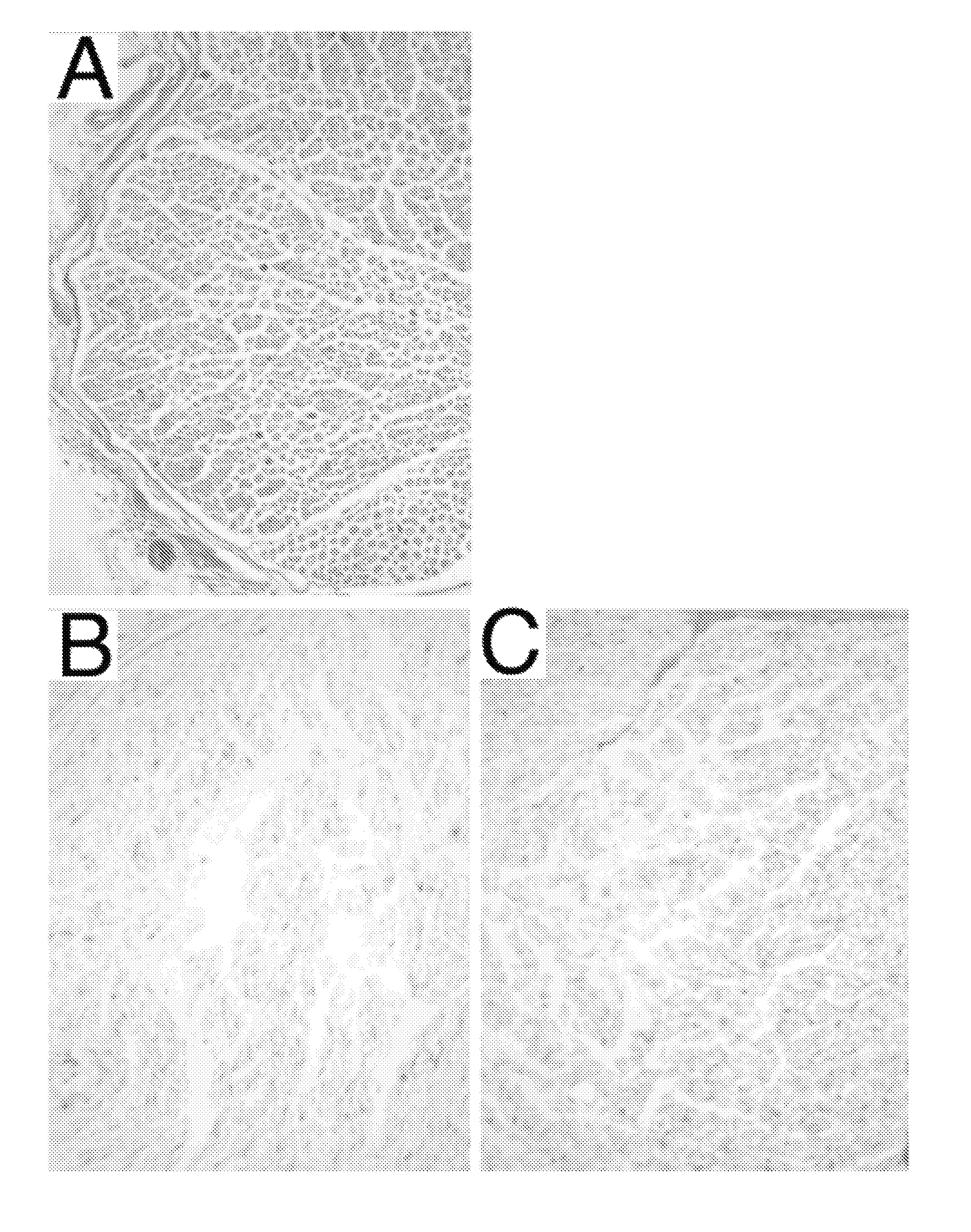
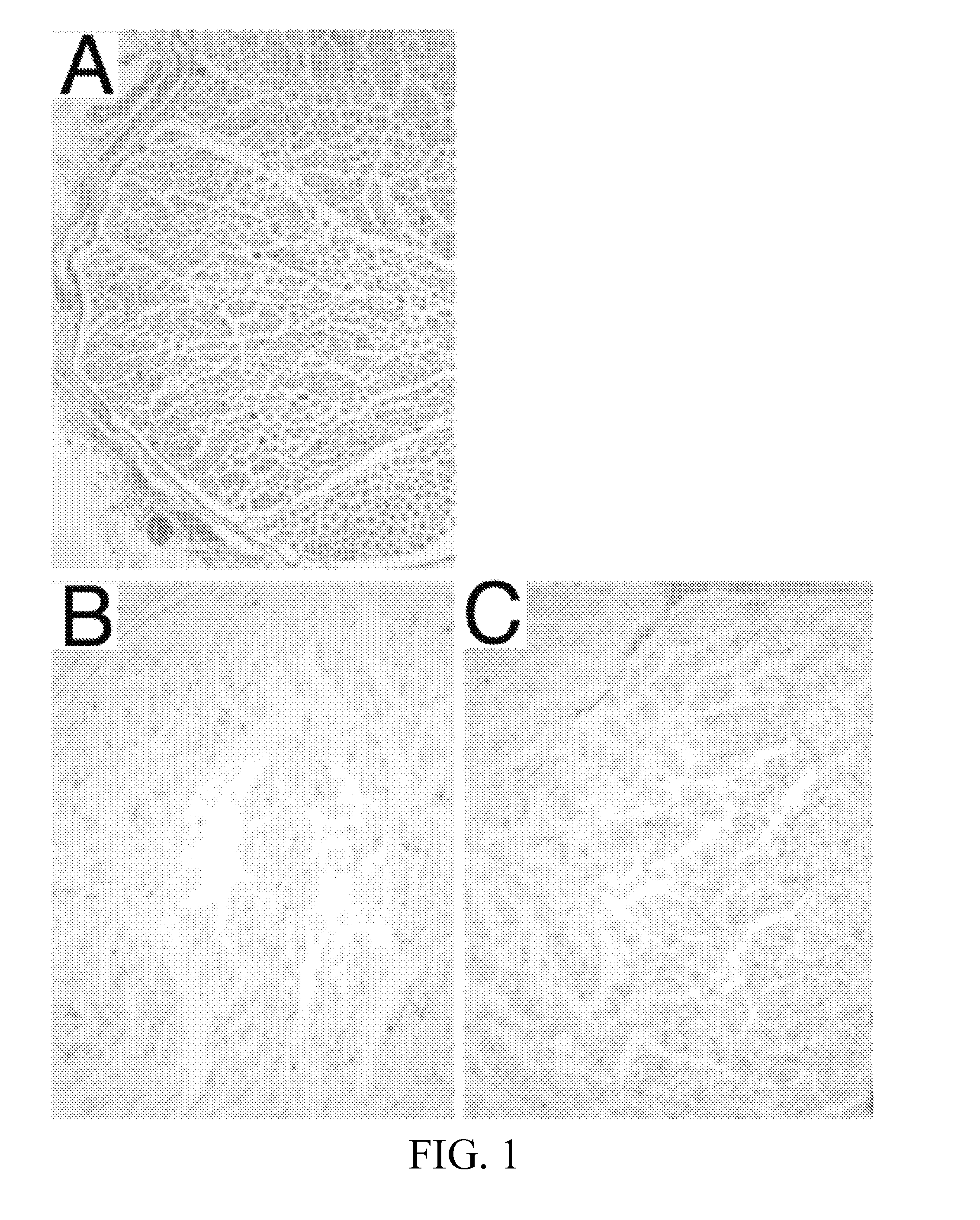
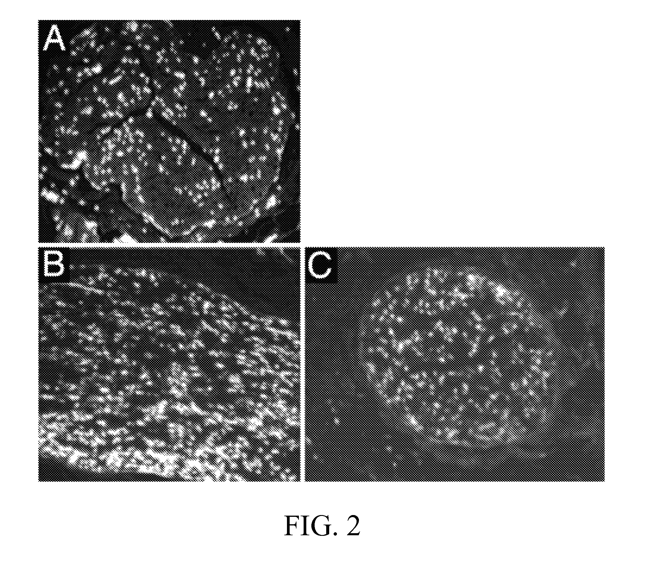
[0086]The DC2 and DC3 grafts from the two processing methods were evaluated by several histological techniques. Results of each evaluation were semi-quantitatively scored (0=Not extracted, 1=Partially extracted, 2=Mostly extracted, 3=Fully extracted and R=Redistributed). Results for the two detergent processing schemes (DC2, DC3) and normal nerve control are shown in FIGS. 1-6.

[0087]FIG. 1 illustrates hematoxylin and eosin (H&E) stains of rabbit nerve tissue (control; panel A), rabbit nerve tissue processed in accordance with the process taught by Hudson et al. (DC2; panel B), and rab...

example 3

Immunocompatibility of Nerve Grafts

[0093]Extensive in vivo testing in New Zealand White (NZW) rabbits has been performed with nerve grafts processed by the Hudson et al. method and the subject invention. Results show that both types of decellularized nerve grafts were immunologically compatible and no graft rejection was observed.

[0094]NZW rabbits are an outbred strain. It is known in the literature that allografting of live or cellular tissues from a NZW rabbit donor to NZW recipient results in graft immunorejection.

[0095]Therefore, the observed lack of graft rejection in NZW allografts demonstrates that nerve allografts prepared by the subject invention are immunocompatible.


PUM

PropertyMeasurementUnit
pHaaaaaaaaaa
temperatureaaaaaaaaaa

Abstract

The subject invention pertains to materials and methods for processing tissue to produce a natural, acellular replacement tissue that is immunocompatible with a recipient. According to the subject invention, harvested tissue is subjected to wash solutions wherein only amphoteric detergent(s) are used (e.g., anionic detergents are excluded). Following extraction by amphoteric detergent(s), the tissue is washed with a buffer system to facilitate the clearance of cellular components and detergent. In one embodiment, the subject invention pertains to a replacement tissue that is a nerve graft that supports axonal regeneration, guides axons toward the distal nerve end and / or is immunologically tolerated. Preferably, the nerve graft retains essential extracellular matrix scaffolding as well as biological components that promote nerve regeneration through the nerve graft.

Description

CROSS-REFERENCE TO A RELATED APPLICATIONS[0001]This application is a National Stage Application of International Application No. PCT / US2014 / 030688, filed Mar. 17, 2014; which claims the benefit of U.S. provisional application Ser. No. 61 / 794,012, filed Mar. 15, 2013, both of which are incorporated herein by reference in their entirety.BACKGROUND OF THE INVENTION[0002]Biomedical engineering faces many challenges in the development of tissue that can be used to facilitate healing. For example, a “replacement” tissue should promote tissue regeneration. In doing so, the new tissue must be compatible with the recipient tissue so that neighboring cells accept the replacement. Importantly, the replacement tissue should overcome (or avoid) the immunologic responses typically elicited by the addition of a “foreign body” to a biological system.[0003]Furthermore, the replacement tissue should exhibit the properties and function of the tissue that it is replacing. For example, the replacement t...

Claims


Application Information

Patent Timeline
Patent Type & Authority Patents(United States)
IPC IPC(8): C12N5/00A61K35/12A61L27/36C12P1/00C12N5/02A61F2/04
CPCA61L27/3687A61L2430/32C12N5/0081C12N2509/00A61L27/3633A61L27/3675A61L27/54A61L2430/40
Inventor MUIR, DAVID F.
Owner UNIV OF FLORIDA RES FOUNDATION INC
Why Eureka
Try Eureka