
Bacterial surface display and screening of thioether-bridge-containing peptides

Active Publication Date: 2017-05-16
APPLIED NANOSYST
View PDF6 Cites 0 Cited by
  • Summary
  • Abstract
  • Description
  • Claims
  • Application Information

AI Technical Summary

Benefits of technology

The patent describes a method for modifying peptides using a special enzyme. This enzyme can add a ring structure to the peptide, making it more stable and increasing its biological activity. The peptide can be any peptide that can be modified by this enzyme, such as hormones, enzyme inhibitors, or antibodies. The method can also be used on lanthibiotics, which are a type of antibiotic. The peptide can be a mutant of a lanthibiotic or a non-lantibiotic bacteriocin. The method can be used to alter the biological activity of the peptide, making it more or less effective in its intended use.

Problems solved by technology

However, the proteolytic susceptibility and instability of these peptides has been recognized as a major disadvantage for therapeutic applications.
However, at present it is still difficult to efficiently introduce such structures into synthetic peptides, especially for large peptides.

Method used


Image

  • Bacterial surface display and screening of thioether-bridge-containing peptides
  • Bacterial surface display and screening of thioether-bridge-containing peptides
  • Bacterial surface display and screening of thioether-bridge-containing peptides

Examples


example 1

Expression Vector for Cell Surface Display of Thioether and Dehydroresidue-Containing Peptides on Lactococcus Lactis

Objective:

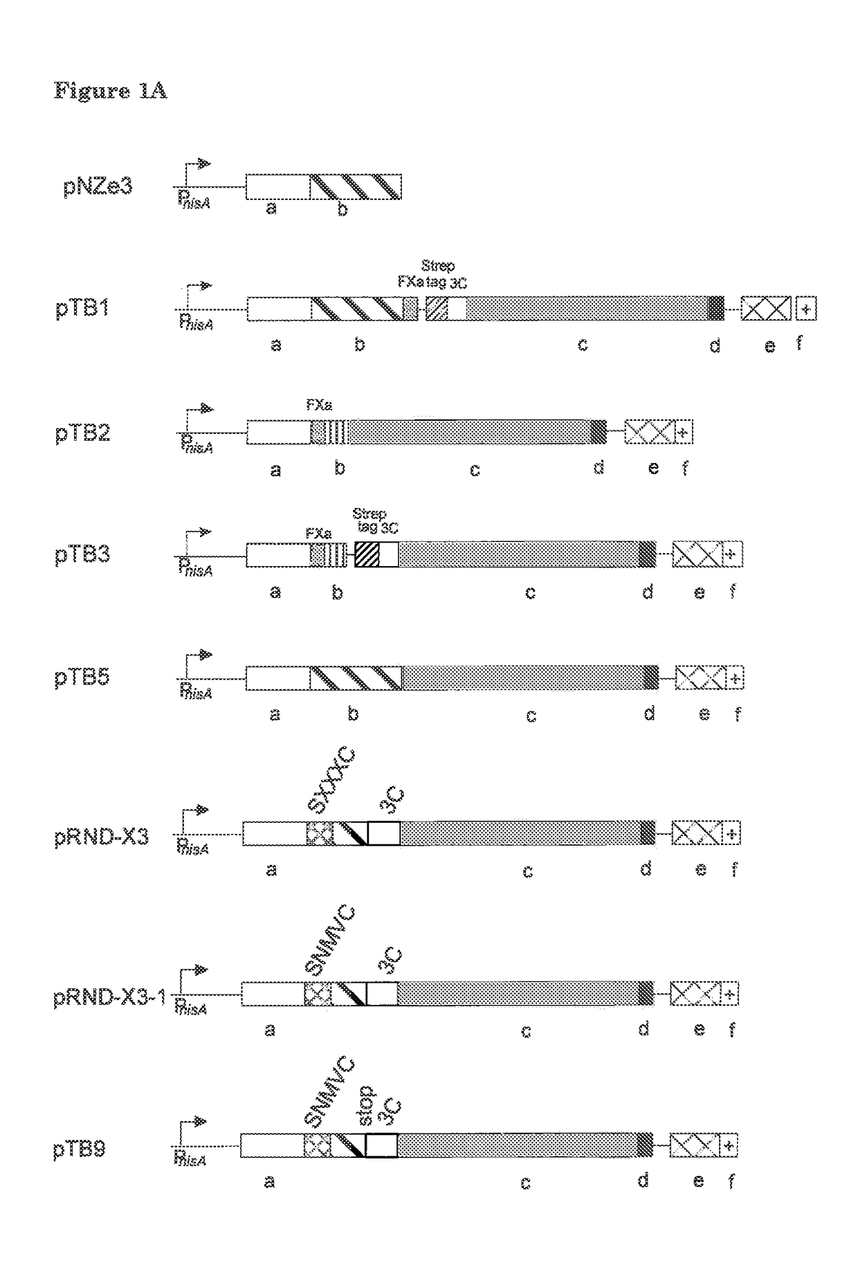
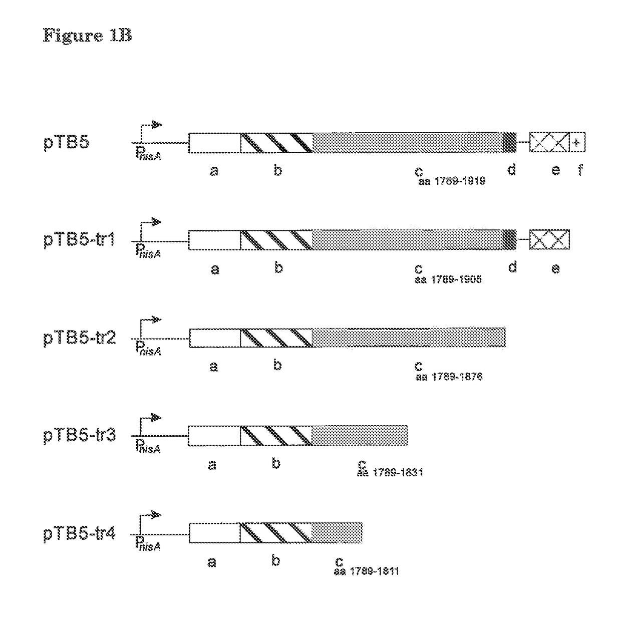
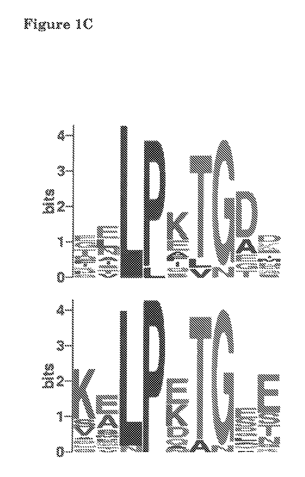
[0054]This example concerns the construction of an expression vector for surface display of thioether-containing peptides on Lactococcus lactis. The L. lactis host organism provides the nisin biosynthesis and export machinery for introduction of the thioether linkages in the desired peptide and its export. The peptide is translationally fused to a LPXTG cell wall-anchoring motif such as that of the L. lactis PrtP protease. This anchoring mechanism requires processing by a sortase for covalent anchoring of the peptide to the peptidoglycan of the bacterial cell wall. In this way the peptide and the encoding DNA are linked allowing selection / screening for post-translationally modified peptides with desired properties. Nisin will be used as a model peptide for development and demonstration of the display system.

Materials and Methods

[0055]The LPXTG cell wall-anch...

example 2

Anti-Leaderpeptide Antibodies Demonstrate the Cell Surface Location of the Cell Wall Attached Prenisin Anchor Fusion Protein TB5

Objective:

[0057]Display of TB5 at the cell surface of L. lactis was evaluated with a whole cell ELISA using anti-nisin leader antibodies.

Materials and Methods

[0058]L. lactis NZ9000(pTB5) and L. lactis NZ9000(pIL3BTC / pTB5) cells were grown with and without nisin for induction of TB5. After production cells were collected by centrifugation, washed three times with phosphate buffered saline, pH 7.4 (PBS). An equal number of cells displaying TB5 are incubated with a 1000-fold diluted rabbit anti-nisin leader antibody solution in a final volume of 1 ml PBS plus 0.5% BSA at room temperature for 1 hour under rotation. After washing three times with PBS displayed TB5 was visualized by incubation with alkaline phosphatase conjugated goat anti-rabbit IgG (1:10000) and p-nitrophenyl phosphate (0.5 mg / ml) as substrate. The absorbance was determined at 410 nm, which is ...

example 3

[0060]Prior to cell surface display the prenisin-anchor fusion protein has been modified intracellularly by NisB and NisC. Modification by NisB- and NisC-modified prenisin-anchor fusion protein was demonstrated by antimicrobial activity against overlaid cells after leader peptide cleavage.

Objective:

[0061]Example 2 shows that TB5 is produced resulting in the display of prenisin on the lactococcal cell surface. In this example the modification of nisin by NisB and NisC was evaluated with an overlay of a nisin-sensitive L. lactis strain. Growth inhibition of this strain indicated that NisB and NisC correctly modified and formed at least ring A, B, and C of TB5.

Materials and Methods

[0062]A GM17 agar plate with an extensively washed SDS-PAA gel or spots of pTB5 producing L. lactis NZ9000 cells was covered with a 200-fold diluted L. lactis MG1363 or NZ9000 strain in 0.5% top agar with 0.1 mg / ml trypsin. Trypsin is required for cleaving of the nisin leader yielding active nisin. The agar p...


PUM

PropertyMeasurementUnit
Fractionaaaaaaaaaa
Electric chargeaaaaaaaaaa
Lengthaaaaaaaaaa

Abstract

The invention relates to bacterial cell surface display of post-translationally modified heterologous proteins. Provided is an isolated nucleic acid construct encoding a proteinaceous substance comprising, from the N-terminus to the C-terminus, at least (a) an N-terminal a lantibiotic leader sequence; (b) an amino acid sequence of interest to be post-translationally modified to a dehydroresidue- or thioether-bridge containing polypeptide; (c) a hydrophilic cell-wall spanning domain; (d) a sortase recognition motif; (e) a hydrophobic membrane spanning domain and (f) a C-terminal charged membrane anchoring domain. Also provided is a Gram-positive host cell expressing the construct, as well as a library of host cells.

Description

[0001]This application is the U.S. National Phase of, and Applicants claim priority from, International Application No. PCT / NL2011 / 050484 filed Jul. 6, 2011 and European Patent Application No. 10168589.9 filed Jul. 6, 2010, each of which are incorporated herein by reference.BACKGROUND OF THE INVENTION[0002]The invention relates to the field of protein engineering and screening for therapeutically relevant peptide. More specifically, it relates to cell surface display of post-translationally modified heterologous proteins. Heterologous display of proteins or peptides on the surface of a micro-organism (e.g. bacteria) is a useful research tool and has been associated with a broad range of interesting applications. Linking the protein or peptide function to the encoding gene enables the selection and / or optimization of peptides with desired properties from large combinatorial libraries. Various display formats have been developed including, ribosome display, phage display, bacterial su...

Claims


Application Information

Patent Timeline
IPC IPC(8): C12N15/10C12N15/74G01N33/68C12N15/62
CPCG01N33/6803C12N15/1037C12N15/625C07K2319/035C07K2319/50
Inventor BOSMA, TJIBBE
Owner APPLIED NANOSYST
Learn More