Renal tissue regeneration

a technology of renal failure and tissue regeneration, applied in the field of tissue engineering, can solve the problems of extremely limited treatment options for renal failure, and achieve the effect of reducing or eliminating the need for donor cell procuremen

Active Publication Date: 2017-07-04
WAKE FOREST UNIV HEALTH SCI INC
View PDF38 Cites 0 Cited by
  • Summary
  • Abstract
  • Description
  • Claims
  • Application Information

AI Technical Summary

Benefits of technology

[0009]This invention reduces or eliminates the need for laborious stem cell isolation and ex vivo cell culturing by directly recruiting a stem cell population into target specific sites for regeneration in vivo. This is possible due to the nature of host cellular responses to environment changes. In one aspect of the invention, the use of a biocompatible substance as an implant can be used to recruit pluripotent cells to a target site within a body structure and induce tissue regeneration within the body structure in vivo using a “directed cell recruitment” technique. Moreover, a method can be directed to inducing cell recruitment to a target site by delivering a biocompatible substance to a target site within a body structure to produce positive pressure within the target site and maintaining the positive pressure, such that pluripotent cells are recruited to the target site. Another aspect can be directed to a method of inducing in situ tissue regeneration at a target site of a subject by delivering a biocompatible substance to the target site to produce a positive pressure, maintaining the positive pressure for a period of time such that the number of pluripotent cells is increased at the target site, delivering at least one adjuvant to the target site and promoting differentiation of the pluripotent cells to regenerate the tissue.
[0016]In another aspect, the invention discloses target specific gel scaffolds. These target specific gel scaffolds can maximize the tissue regenerative capacity. Incoporation of factors, such as growth factors, extracellular matrix (ECM) proteins and bioactive molecules into the gel scaffold system can enhance tissue formation. This gel system can be used as a preventive measure in subjects with high risk of organ or tissue failure. The scaffolds can take various forms, depending on the tissue and its use. The biocompatible substance can be delivered into the target site via injection, surgically placing the substance into the target site, or by using a catheter.

Problems solved by technology

Current treatment options for renal failure are extremely limited and only renal transplantation can restore kidney function.

Method used


Image

  • Renal tissue regeneration
  • Renal tissue regeneration
  • Renal tissue regeneration

Examples


example 1

nd Materials

Normal Mouse Model

[0096]All animal procedures were performed in accordance with a protocol approved by the institutional Animal Care and Use Committee (ACUC) at Wake Forest University. CD1 mice (6-8 weeks) were purchased from Charles River Laboratories Inc. (Wilmington, Mass.). Under anesthesia using isoflurane, the kidneys were accessed through a dorsal incision and then collagen gels were injected into kidneys. Mice were divided into three experimental groups (n=20 per time point). The following treatments were administered via multiple injections into both kidneys using a 22-gauge needle: (1) collagen gel (0.2% wt / vol, 50 μL / kidney), (2) saline (0.9% NaCl, 50 μL / kidney, Hospira, Inc, Lake Forest, Ill.), and (3) needle sticks only. Sham operation served as a control. The kidneys were harvested at 1, 2, 3, and 4 weeks after injection for histological and immunohistochemical analyses.

Collagen Gel Preparation

[0097]Rat tail type I collagen solution was obtained from BD Bio...

example 2

Implantation

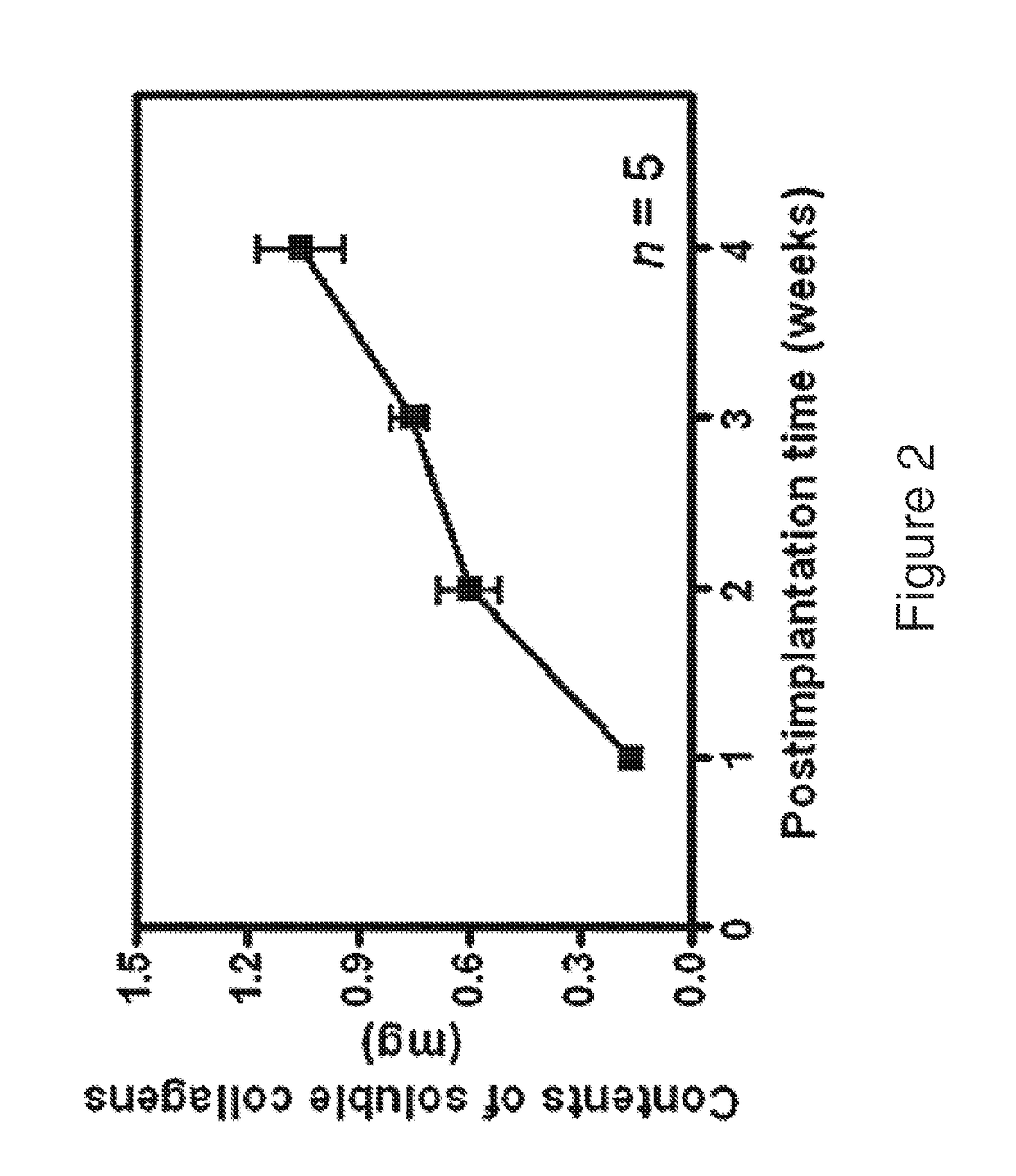
[0129]The PGA nonwoven scaffolds consisted of highly porous fiber mesh disks. The retrieved scaffolds at 1, 2, 3 and 4 weeks showed a progressive tissue ingrowth over time. Cell recruitment after implantation was measured by gross morphology and SEM microphotograph. By the fourth week, the implants were completely encapsulated by a connective tissue capsule with abundant host vasculature. Neovascularization was observed in and around the scaffold. Examination of the retrieved implants with scanning electron microscopy (SEM) clearly demonstrated a classic foreign body reaction. The porous scaffold became populated with collagen-producing cells and the interfiber spaces were filled with increasing amounts of collagenous matrix when measured by SEM microphotographs each week after implantation for 4 weeks. Observations after implantation, noted extracellular matrix material gradually filled in the porous architecture of the scaffold.

example 3

Component Measurement

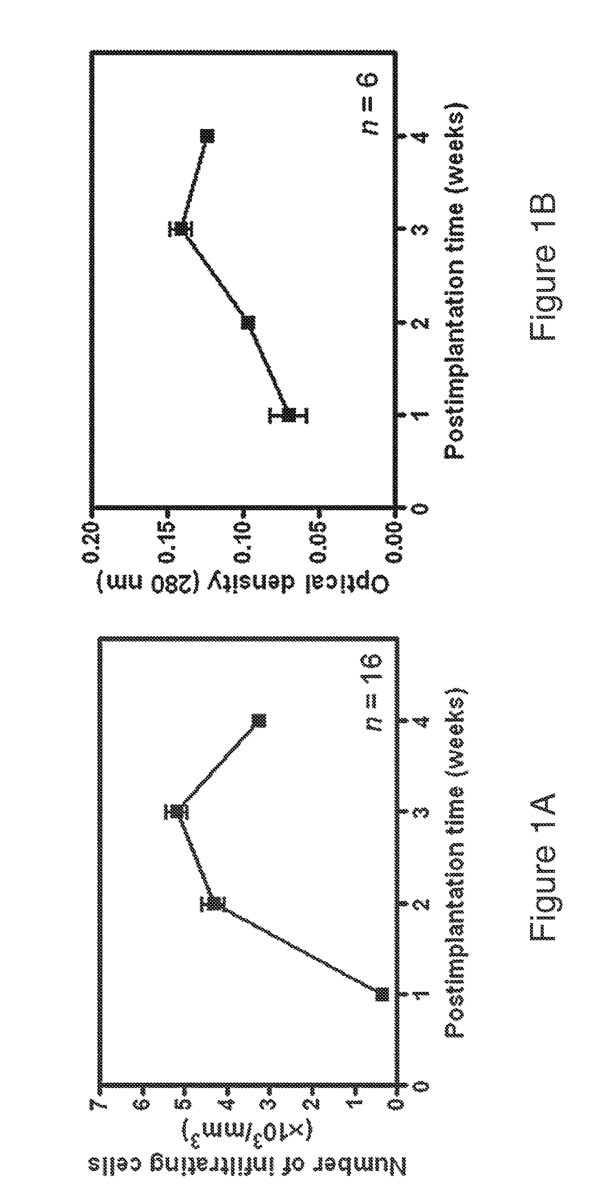
[0130]Measurement of cell density within the scaffold was achieved by histomorphometry and DNA content analyses (FIG. 1). DNA analysis, performed using a standard extraction kit, confirmed the typical host cellular response. Cell infiltration increased steadily in the first three weeks, followed by a slight decline as matrix production increased by the fourth week. These results were confirmed by histomorphometry, where the average cell counts of representative H&E stained sections showed a similar pattern.


PUM

PropertyMeasurementUnit
concentrationaaaaaaaaaa
viscosityaaaaaaaaaa
pressureaaaaaaaaaa

Abstract

The invention is directed to methods of inducing cell recruitment and tissue regeneration at a target site in a subject. It is also based, in part, on the discovery that a subject's own biologic resources and environmental conditions can be used for in situ tissue regeneration and thereby reduce or eliminate the need for donor cell procurement and ex vivo manipulation of such donor cells. Methods are disclosed for recruitment of a subject's own stem cells to a target region by inducing a sustained positive pressure at a target site, such as the kidney, thereby increasing the number of pluripotent cells capable of differentiating to regenerate the target tissue.

Description

FIELD OF THE INVENTION[0001]The present invention concerns tissue engineering and, in particular, methods of recruiting stem cells to a target site.BACKGROUND OF THE INVENTION[0002]Chronic renal disease is a common condition that elevates the risk of complete renal failure, cardiovascular disease and other complications. The prevalence of chronic renal failure has continuously increased in the United States in the last decade. Currently, the only treatment options for renal failure are dialysis and transplantation, both of which are associated with considerable cost. Dialysis, usually performed 3 times per week, impairs the patient's quality of life and does not replace renal functions such as synthesis of erythropoietin and vitamin D. Transplantation, on the other hand, can replace all renal functions, but the rising occurrence of end stage renal disease (ESRD) in the United States continues to outpace the rate of organ donation, as reflected by the fact that the waiting list conti...

Claims


Application Information

Patent Timeline
Patent Type & Authority Patents(United States)
IPC IPC(8): A61K47/30A61K9/00A61K38/39A61K38/48A61L27/54A61L27/24A61L27/52
CPCA61K38/39A61K9/0024A61K38/4886A61L27/24A61L27/52A61L27/54A61L2300/41A61L2300/434A61L2400/06A61L2430/26A61K2300/00A61P13/12
Inventor YOO, JAMESLEE, SANG JINVAN DYKE, MARKATALA, ANTHONY
Owner WAKE FOREST UNIV HEALTH SCI INC
Why Eureka
Try Eureka