
Bone and metal targeted polymeric nanoparticles

a polymer and nanoparticle technology, applied in the field of nanoparticles, can solve the problems of increasing the risk of fracture, so as to increase the rate of osseointegration, enhance the osseous integration of the implant, and reduce the risk of fractur

Active Publication Date: 2018-01-23
THE BRIGHAM & WOMEN S HOSPITAL INC +1
View PDF3 Cites 0 Cited by
  • Summary
  • Abstract
  • Description
  • Claims
  • Application Information

AI Technical Summary

Benefits of technology

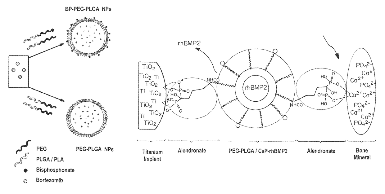
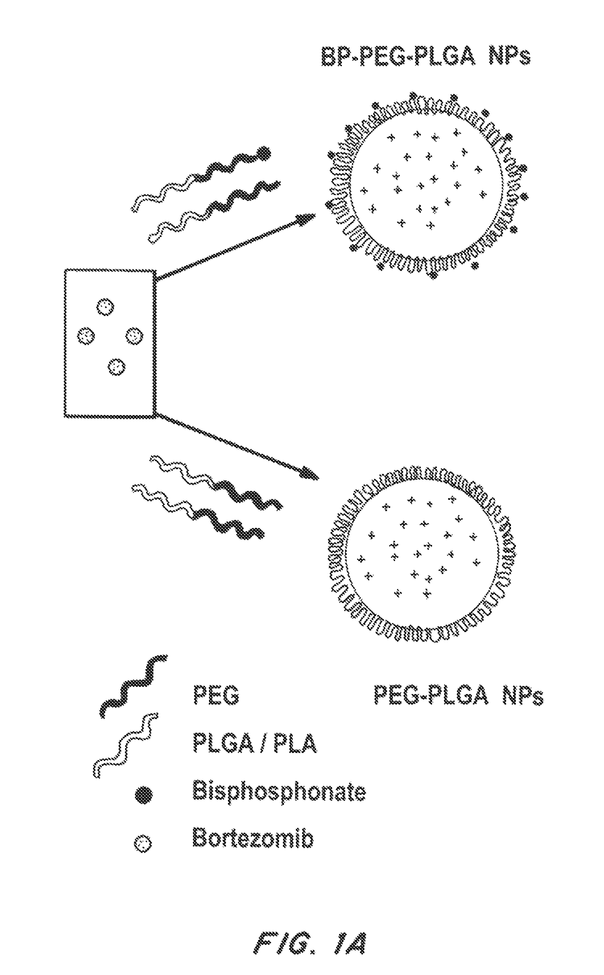
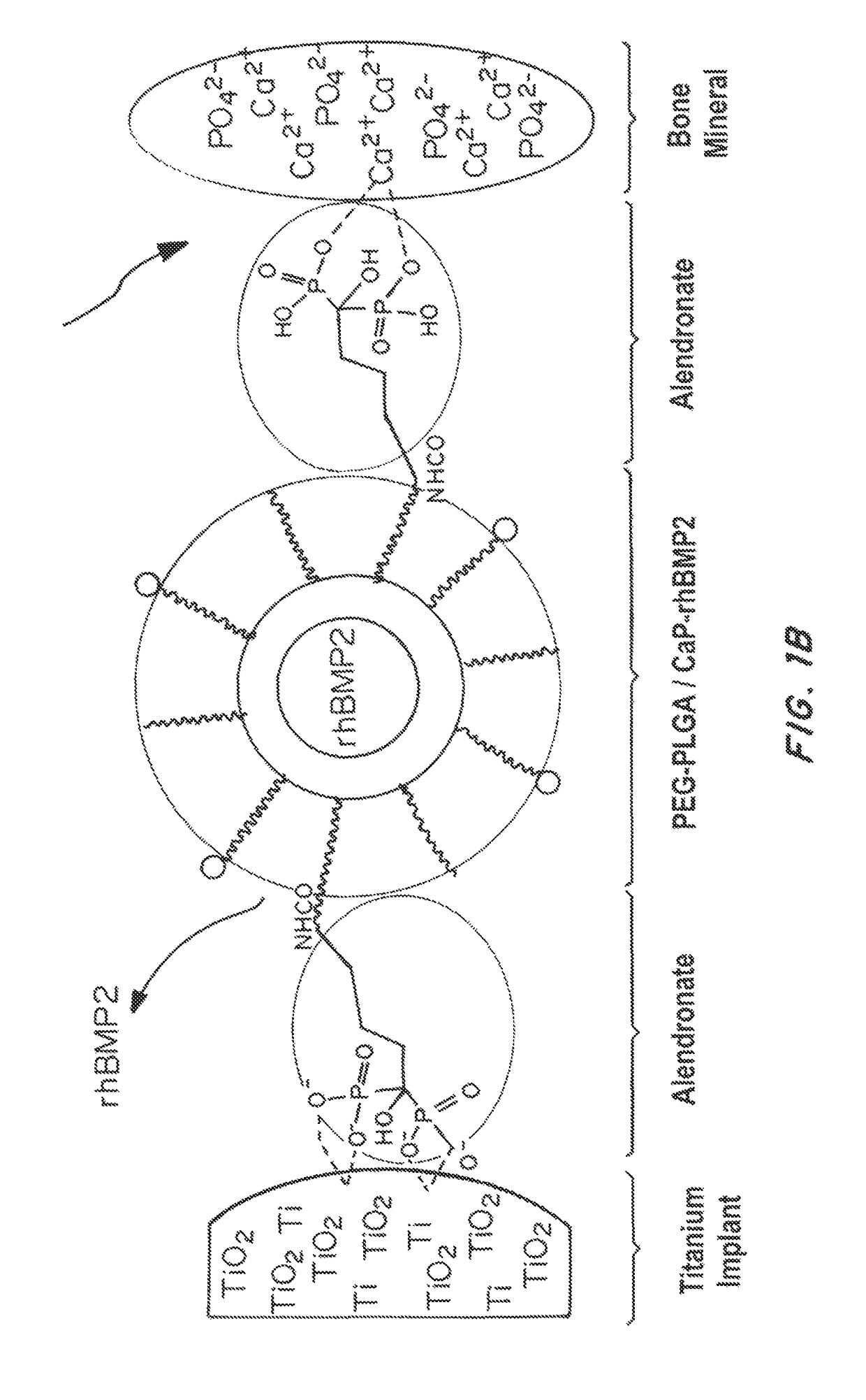
This patent describes a method of creating polymeric nanoparticles that can target bone and metal surfaces, and can carry therapeutic agents for local delivery. These nanoparticles have a targeting element that binds to bone and minerals, a "stealth layer" to evade immune response, and a biodegradable polymer inner core. By coating surgical implants with these nanoparticles, they can increase the rate of bone growth and decrease infection. The nanoparticles adhere easily to metal implants, allowing for quick and controlled delivery of drugs to the implant-tissue interface during surgery. This patent provides a novel method for improving the healing and integration of surgical implants.

Problems solved by technology

When cancer metastasizes to bone, it becomes almost incurable and creates considerable pain.
However, these strategies are limited by the harsh conditions and time-consuming procedures for coating the device, and offer little for practical applications.
There has been literally no practical solution when it comes to intraoperative modification procedure of the surgical implants for local drug delivery of therapeutics such as antibiotics, growth factors etc., for individualized patient treatment.
There is a complete absence of any method that can provide flexibility of modifying surgical implants during an operation or in emergencies.
There is no effective strategy for local drug delivery at the surgical metal implant-bone tissue interface that can enhance osseointegration of implants in the tissue or that can actively promote the tissue growth at the implant surface.

Method used


Image

  • Bone and metal targeted polymeric nanoparticles
  • Bone and metal targeted polymeric nanoparticles
  • Bone and metal targeted polymeric nanoparticles

Examples


example 1

of PLGA-PEG-COOH and PLGA-PEG-Alendronate

[0160]Materials and Methods:

[0161]Reagents

[0162]The polymer Poly(D,L-lactide-co-glycolide) (50 / 50) with terminal carboxylate groups (PLGA, inherent viscosity 0.26-0.54 dl / g in hexafluoroisopropanol, MW 25 kDa) was obtained from Lactel Absorbable Polymers, USA. NH2-PEG-COOH (MW 3400) and BOC—NH2-PEG-COOH (MW 3400) were purchased from Laysan Bio, USA. All reagents were analytical grade or above and used as received, unless otherwise stated. The recombinant bone morphogenic protein (BMP) and BMP-2 Elisa kit was procured from R&D systems. Alkaline assay kit was purchased from LP assay Kit abCam. The alamar blue reagents, and Cy3-COOH, Alexa-NH2 dyes (647, 488, 405) was purchased by Invitrogen. The sodium alendronate, ascorbic acid, beta-gycerol phosphate, N-Hydroxysuccinimide (NHS), Ethyl-3-(3-dimethylaminopropyl) carbodiimide (EDC), hydroxy apatite, Alizarine red and any other chemicals were purchased from Sigma Aldrich. Bortezomib was purchased...

example 2

on of Bone Targeted and Non-Targeted PLGA-b-PEG-Ald and PLGA-b-PEG-Ald / CaP NPs

[0166]Materials and Methods

[0167]Nanoparticles (NPs) were prepared by nanoprecipitation and emulsion methods. In brief, Ald-PEG-PLGA (10 mg / ml, in acetonitrile) was mixed with therapeutic molecules (in DMSO) just before NP formation. NPs were formed by adding polymer-drug mixture dropwise into water to give a final concentration of 1 mg / ml. NPs were stirred for 4 hrs.

[0168]In the single emulsion method of Ald-PEG-PLGA NP formulation, 5 mg / ml of polymer blend of PLGA-PEG-BP to PLGA-PEG in a weight ratio of 1:5 (in chloroform) is mixed with a chemotherapeutic drug (e.g., Bortezomib) and emulsified with aqueous phase, using a probe sonicator for 90 to 120 seconds at 50 watt power for with pulses of 5 second on and intermediate 1 second stop time. The organic to aqueous phase volume ratio was between, 1:8 to 1:15. The emulsions were stabilized by adding polyvinyl alcohol (0.2%) to the aqueous phase. The NP emu...

example 3

Alendronate to Polymer in the NPs

[0179]Materials and Methods

[0180]The ratio of alendronate to polymer in the NPs was determined by phosphate assay. The PLGA-b-PEG-Ald NPs (10 μl of 10 mg / ml NPs in water) were mixed with 60 μl of concentrated H2SO4 and 10 μl of H2O2 in glass tubes and was heated for 10 minutes at 200° C. Thereafter, 690 μl Na2S2O5 (3 mg / ml) was added to the tubes and the samples were incubated at 100° C. for 5 minutes. This was followed by adding 200 μl of (NH4)6Mo7O24.4H2O (20 mg / ml) and 20 μl of ascorbic acid (100 mg / ml) to the glass tubes and incubate for another 10 minutes at 100° C. The absorbance of the sample solutions were determined at 820 nm and were used to determine the phosphate concentrations of the samples. The calibration curves were based on known concentrations of Na2HPO4 as standards. Non-targeted PLGA-PEG NPs were used as negative control.

[0181]Results

[0182]FIG. 4 shows the ratio of alendronate / PEG-PLGA in pre and post formulation conjugation. The...


PUM

PropertyMeasurementUnit
diameteraaaaaaaaaa
diameteraaaaaaaaaa
diameteraaaaaaaaaa

Abstract

Bone- and metal-targeted polymeric nanoparticles are provided. Exemplary nanoparticles have three main components: 1) a targeting element that can selectively bind to bone, minerals, or metal ions; 2) a layer of stealth to allow the polymer to evade immune response; and 3) a biodegradable polymeric material, forming an inner core which can carry therapeutics or other diagnostics. Preferred nanoparticles contain a blend of target-element polymer conjugate and polymer that optimizes the ligand density on the surface of the nanoparticle to provide improved targeting of the nanoparticle. The ratio of target-element polymer conjugate to polymer can also be optimized to improve the half-life of the nanoparticles in the blood of the subject. The nanoparticles also exhibit prolonged, sustained release of therapeutic agents loaded into the particles.

Description

CROSS-REFERENCE TO RELATED APPLICATIONS[0001]This application claims the benefit of priority to U.S. Ser. No. 61 / 899,089, filed on Nov. 1, 2013, the disclosure of which is incorporated herein by reference.FIELD OF THE INVENTION[0002]This invention is generally directed to the field of nanoparticles including nanoparticles for drug delivery.BACKGROUND OF THE INVENTION[0003]Three-fourths of the patients with breast and prostate cancer develop complications of bone metastasis. When cancer metastasizes to bone, it becomes almost incurable and creates considerable pain. Metastasizing cancer also deregulates bone remodeling, leading to bone fractures, hypercalcaemia, spinal cord and nerve compression syndromes.[0004]The mineralized extracellular matrix and specific cell types in the bone microenvironment are controlled by local and systemic factors that create a special milieu to provide a fertile soil for many cancers in which to thrive, including metastasizing cancer from other distant ...

Claims


Application Information

Patent Timeline
Patent Type & Authority Patents(United States)
IPC IPC(8): A61K9/00A61K38/18A61K38/05
CPCA61K47/6935A61K47/6937A61K47/6957A61K38/05A61K38/1875A61K47/548
Inventor SWAMI, ARCHANABASTO, PAMELAKARP, JEFFREYFAROKHZAD, OMID C.
Owner THE BRIGHAM & WOMEN S HOSPITAL INC
Learn More