Methods for detection of nucleic acid sequences in urine

a nucleic acid sequence and urine technology, applied in the field of non-invasive methods, can solve the problems of ineffective methods, inconvenient diagnosis of certain genetic defects, and intrusion of genetic material for diagnosis in certain cases, and achieve the effect of reducing dna degradation

Inactive Publication Date: 2007-11-13
TROVAGENE
View PDF11 Cites 51 Cited by
  • Summary
  • Abstract
  • Description
  • Claims
  • Application Information

AI Technical Summary

Benefits of technology

[0042]It is helpful in some embodiments to include a step to reduce DNA degradation in said urine sample, which in one embodiment encompasses treatment with a compound selected from the group comprising: ethylenediaminetetraacetic acid, guanidine-HCl, Guanidine isothiocyanate, N-lauroylsarcosine, and Na-dodecylsulphate.

Problems solved by technology

Until now, the fragile nature of nucleic acids, and their location encapsulated within cells, made the acquisition of genetic material for diagnosis in certain cases necessarily intrusive.
Such intrusive practices carry with them a level of risk to both the fetus and the mother.
While developments in ultrasound have contributed less intrusive alternative methods of fetal monitoring during pregnancy, these methods are not appropriate for diagnosing certain genetic defects and are not effective during the early stages of pregnancy, even for determining fetal sex.
However, the fate of these chromatin degradation products in the organism has not been investigated in detail.
Necrosis is considered to be a catastrophic metabolic failure resulting directly from severe molecular and / or structural damage.
Available data on the fate of these chromatin degradation products in organisms provide little guidance.
It can be difficult to ensure that this DNA did not originate from white blood cells as a result of their lysis during sample treatment.
U.S. Pat. No. 5,496,699, to George D. Sorenson. However, the use of blood or plasma as a source of DNA is both intrusive to the patient and problematic for the diagnostic technician.
In particular, a high concentration of proteins (about 100 mg / ml) as well as the presence of compounds which inhibit the polymerase chain reaction (PCR) make DNA isolation and analysis difficult.

Method used


Image

  • Methods for detection of nucleic acid sequences in urine
  • Methods for detection of nucleic acid sequences in urine
  • Methods for detection of nucleic acid sequences in urine

Examples


example 1

DETECTION OF POLYMERIC DNA IN URINE OF MICE PREINOCULATED WITH λ PHAGE DNA

[0161]This example analyzes the ability of DNA to cross the kidney barrier in rodents and appear in detectable form in urine.

[0162]λ phage DNA (New England BioLabs, MA) was labeled by nick translation with [α-32P] dNTP DNA (New England BioLabs, MA) using the Klenow fragment of E. Coli DNA polymerase to a specific radioactivity of 108 cpm / μg as previously described. ( / ]Sambrook J., Fritsch E. F., Maniatis T., Molecular Cloning. A Laboratory Manual. 2d Edition. Cold Spring Harbor Laboratory Press, 1989). Two months old male Wistar rats were injected subcutaneously with 0.4 μg of the [32p] labeled DNA. Urine samples were then collected for three days and total and acid-insoluble radioactivity was measured in a liquid scintillation counter. The kinetics of excretion of acid-insoluble radioactivity in urine appear in Table 1, below. It was determined that 3.2% of the total DNA with which the rats were inoculated cr...

example 2

DETECTION OF HUMAN DNA SEQUENCES IN URINE OF MOUSE PREINOCULATED WITH HUMAN CELLS

[0167]Example 1 showed that DNA sequences could remain in polymeric form in the blood stream, cross the kidney barrier, and remain suitable for subsequent detection. The next set of experiments were performed to determine if DNA from cells dying in the organism but not in the urinary tract can be detected in urine.

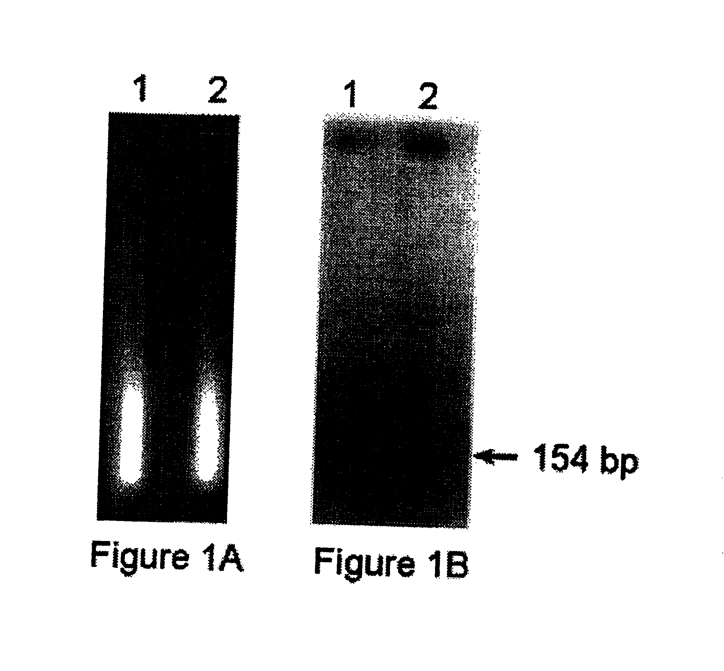
[0168]Human Raji lymphoma cells growing in RPMI supplemented with 5% fetal calf serum were irradiated with 1000 rads of 137CS γ-rays. Mice were then inoculated subcutaneously with 108 cells each. Urine samples were collected for three days, and DNA was isolated as described above. Human-specific DNA sequences were detected by multilocus screening using Alu oligonucleotide-directed PCR as previously described, (Zietkiewicz, E., Labuda M., Sinnett D., Glorieux F. H., and Labuda D., Proc. Natl. Acad. Sci. USA, 89, 8448-8451, 1992) followed by electrophoresis in a 1.5% agarose gel.

[0169]The result...

example 3

DETECTION OF TRANSRENAL NUCLEIC ACIDS

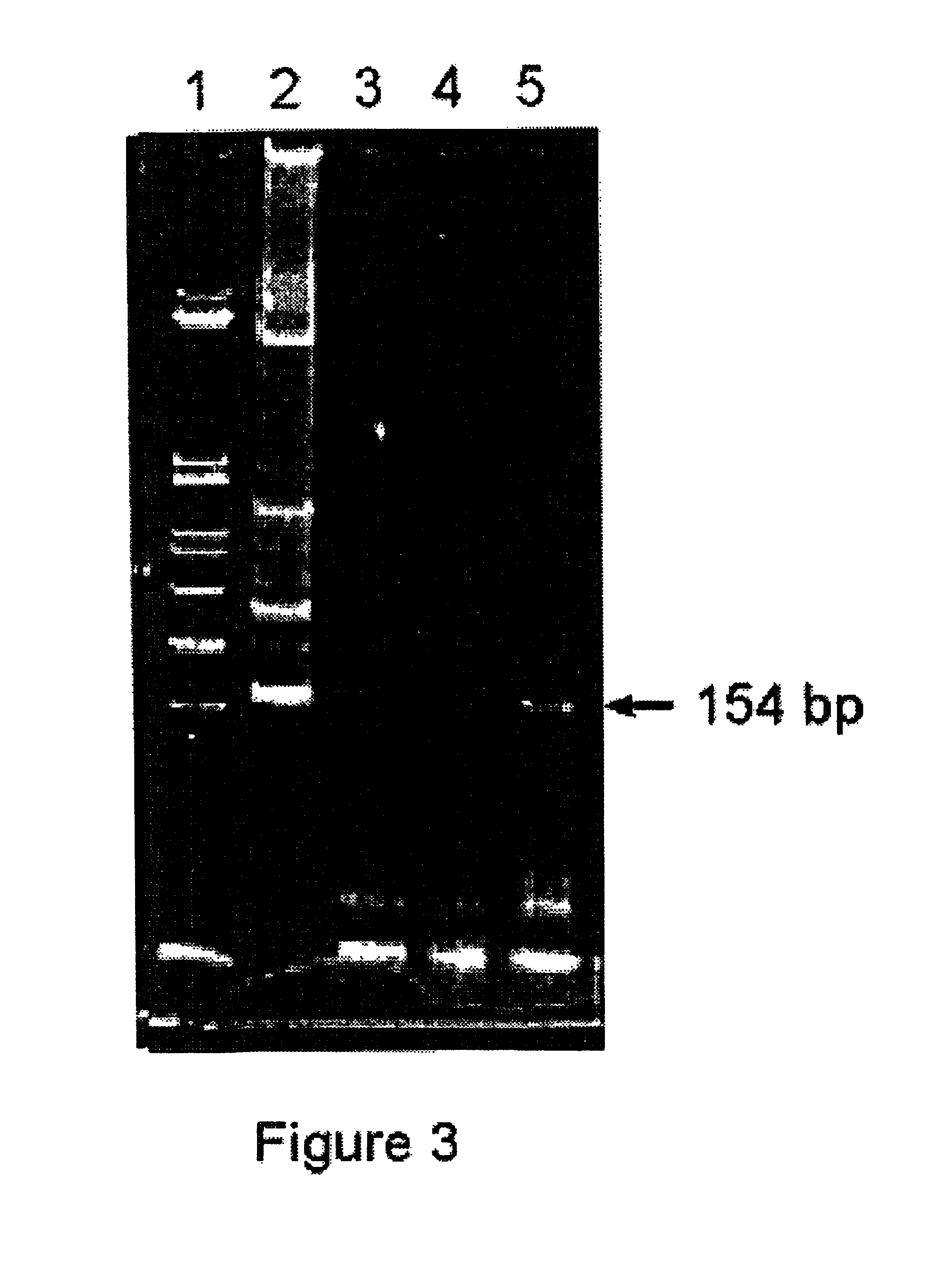
[0171]Taken together, Examples 1 and 2 demonstrate that, in the mouse model, both free DNA and DNA from dying cells cross the kidney barrier and can be detected in urine by PCR analysis. Two systems were selected as models to demonstrate that DNA can cross the kidney barrier and remain in polymeric form in human urine samples. The systems are designed to focus on DNA originating from cells dying outside the urinary tract rather than DNA that appears in urine due to the death of cells in the urinary tract.

[0172]Women who were either pregnant or transfused with male blood were studied because both of these models represented humans having DNA in their bodies that is not present in their normal genome. Each woman studied was analyzed for the presence of Y chromosome-specific sequences in their urine.

Detection of repeated and Single Copy Y Chromosome Specific Sequences in the Urine of Women Pregnant with Male Fetuses.

[0173]As discussed above, apoptot...


PUM

PropertyMeasurementUnit
concentrationaaaaaaaaaa
pHaaaaaaaaaa
Tmaaaaaaaaaa

Abstract

Described are non-invasive methods of detecting the presence of specific nucleic acid sequences as well as nucleic acid modifications and alterations by analyzing urine samples for the presence of transrenal nucleic acids. More specifically, the present invention encompasses methods of detecting specific fetal nucleic acid sequences and fetal sequences that contained modified nucleotides by analyzing maternal urine for the presence of fetal nucleic acids. The invention further encompasses methods of detecting specific nucleic acid modifications for the diagnosis of disease, such as cancer and pathogen infections, and detection of genetic predisposition to various disease. The invention specifically encompasses methods of analyzing specific nucleic acid modifications for the monitoring of cancer treatment. The invention further encompasses methods of analyzing specific nucleic acids in urine to track the success of transplanted cells, tissues and organs. The invention also encompasses methods for evaluating the effects of environmental factors and aging on the genome.

Description

CROSS-REFERENCE TO RELATED APPLICATIONS[0001]This application is a continuation-in-part and a division of application Ser. No. 09 / 609,162 filed Jul. 3, 2000, now U.S. Pat. No. 6,287,820, which is a continuation-in-part and a division of application Ser. No. 09 / 230,704, filed Feb. 4, 2000, now U.S. Pat. No. 6,251,704, which is the United States national phase of International Patent Application No. PCT / US98 / 10965, filed May 29, 1998, which claims priority of U.S. provisional patent applications No. 60 / 058,170, filed May 30, 1997, and Ser. No. 60 / 048,381, filed Jun. 3, 1997.STATEMENT OF RIGHTS TO INVENTIONS MADE UNDER FEDERALLY SPONSORED RESEARCH[0002](Not Applicable)TECHNICAL FIELD[0003]The present invention encompasses non-invasive methods of detecting the presence of specific nucleic acid sequences as well as nucleic acid modifications and alterations by analyzing urine samples for the presence of transrenal nucleic acids. More specifically, the present invention encompasses method...

Claims


Application Information

Patent Timeline
Patent Type & Authority Patents(United States)
IPC IPC(8): C12P19/34C07H21/02C07H21/04C12Q1/68G01N33/00
CPCC12Q1/6806C12Q1/6879C12Q1/6883C12Q1/6886Y10T436/143333C12Q2527/125C12Q2525/125C12Q2600/154C12Q2600/156
Inventor UMANSKY, SAMUIL R.LICHTENSTEIN, ANATOLY V.MELKONYAN, HOVSEP S.
Owner TROVAGENE
Why Eureka
Try Eureka