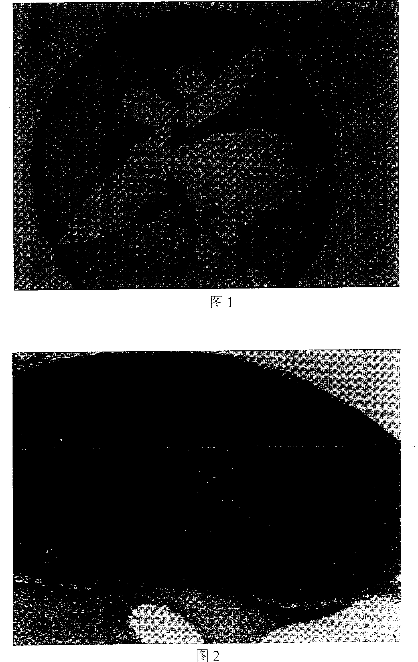
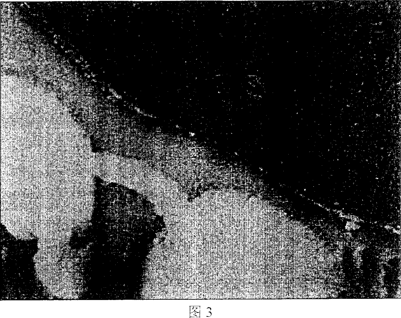
Tissue section cutting method for sea water left eye floundre and right eye founder fertilized egg
A technology for tissue slicing and fertilized eggs, which is applied in the preparation of test samples, instruments, teaching models, etc., to achieve the effect of avoiding slicing failure
- Summary
- Abstract
- Description
- Claims
- Application Information
AI Technical Summary
Problems solved by technology
Method used
Image
Examples
Embodiment 1
[0025] Example 1: Flounder fertilized egg tissue section
[0026] Step 1: Put the flounder fertilized eggs of different developmental stages into 50 times the volume of Bouin's fixative for 10 hours, wash with 70% ethanol for several times (wash away the fixative) and store.
[0027] Step 2: After washing the preserved eggs with 70% ethanol, use a dissection needle to puncture the egg membrane in the direction of the plant pole.
[0028] Step 3: Place it on a glass slide, drip hot-melt agar, and adjust the position and orientation of the eggs under a dissecting microscope.
[0029] Step 4: The embedded agar block is dehydrated by 70%, 80%, 90%, 95%, 100% ethanol as a tissue block, and the treatment time of each group is 30min. A small amount of eosin was added to 95% ethanol to stain the oocytes red.
[0030] Step 5: Put it into terpineol for transparent treatment, and the treatment time is 4 hours.
[0031] Step 6: Put it into a mixed solution of terpineol and hot-melt par...
Embodiment 2
[0035] Example 2: Turbot fertilized egg tissue section
[0036] Step 1: Put the turbot fertilized eggs of different developmental stages in 50 times the volume of Bouin's fixative for 12 hours, wash with 70% ethanol for several times (wash away the fixative) and store.
[0037] Step 2: After washing the preserved eggs with 70% ethanol, use a dissection needle to puncture the egg membrane in the direction of the plant pole.
[0038] Step 3: Place it on a glass slide, drip hot melt agar, and adjust the position and direction of the eggs under a dissecting microscope.
[0039] Step 4: The embedded agar block was dehydrated by 70%, 80%, 90%, 95%, 100% ethanol as a tissue block, and the treatment time for each group was 1 hour. A small amount of eosin was added to 95% ethanol to stain the oocytes red.
[0040] Step 5: Put the dehydrated agar blocks into terpineol for transparent treatment, and the treatment time is 6 hours.
[0041] Step 6: Put it into a mixed solution of terpin...
Embodiment 3
[0045] Example 3: Flounder fertilized egg tissue section
[0046] Step 1: Put flounder fertilized eggs of different developmental stages in 100 times the volume of Bouin's fixative for 6 hours, wash with 70% ethanol for several times (wash away the fixative) and store.
[0047] Step 2: After washing the preserved eggs with 70% ethanol, use a dissection needle to puncture the egg membrane in the direction of the plant pole.
[0048] Step 3: Place it on a glass slide, drip hot melt agar, and adjust the position and direction of the eggs under a dissecting microscope.
[0049] Step 4: The embedded agar block is dehydrated by 70%, 80%, 90%, 95%, 100% ethanol as a tissue block, and the treatment time of each group is 45min. A small amount of eosin was added to 95% ethanol to stain the oocytes red.
[0050] Step 5: Put the dehydrated agar blocks into terpineol for transparent treatment, and the treatment time is 12 hours.
[0051] Step 6: Put it into a mixed solution of terpineol...
PUM
 Abstract
Description
Claims
Application Information
 - R&D
- Intellectual Property
- Life Sciences
- Materials
- Tech Scout
- Unparalleled Data Quality
- Higher Quality Content
- 60% Fewer Hallucinations
Browse by: Latest US Patents, China's latest patents, Technical Efficacy Thesaurus, Application Domain, Technology Topic, Popular Technical Reports.
© 2025 PatSnap. All rights reserved.Legal|Privacy policy|Modern Slavery Act Transparency Statement|Sitemap|About US| Contact US: help@patsnap.com


