Self-controlled laser surgery equipment

A technology of laser surgery and equipment, applied in laser surgery, surgery, ophthalmic surgery, etc., can solve problems affecting the treatment effect, filter bleb rupture, cataract and infection, etc., and achieve the effect of improving the treatment effect and preventing excessive damage

Inactive Publication Date: 2010-03-17
TIANJIN MEDICAL UNIV
View PDF4 Cites 0 Cited by
  • Summary
  • Abstract
  • Description
  • Claims
  • Application Information

AI Technical Summary

Problems solved by technology

Traditional filtering surgery mainly includes trabeculectomy and scleral bite, but there are many complications after traditional filtering surgery, such as early postoperative shallow anterior chamber and no anterior chamber, ciliary prolapse, malignant Glaucoma, hemorrhage, and non-formation of filter blebs, etc.; late stage filter orifice blockage, scar formation, cystoid degeneration, filter bleb rupture, cataract, infection, etc.
The existing laser microsurgery system mainly observes the changes of the biological tissue irradiated by the laser in real time through the microscopic system, and manually controls the amount of laser irradiation during the operation according to the needs of the operation. Therefore, these treatment equipment and treatment methods must observe the operation situation while manually controlling the amount of laser irradiation during the entire operation process, which is cumbersome. Most surgeons rely on their surgical experience to perform laser surgery. It is difficult to control the operation well, and there are still many problems to be solved urgently

Method used


Image

  • Self-controlled laser surgery equipment
  • Self-controlled laser surgery equipment
  • Self-controlled laser surgery equipment

Examples


Embodiment 1

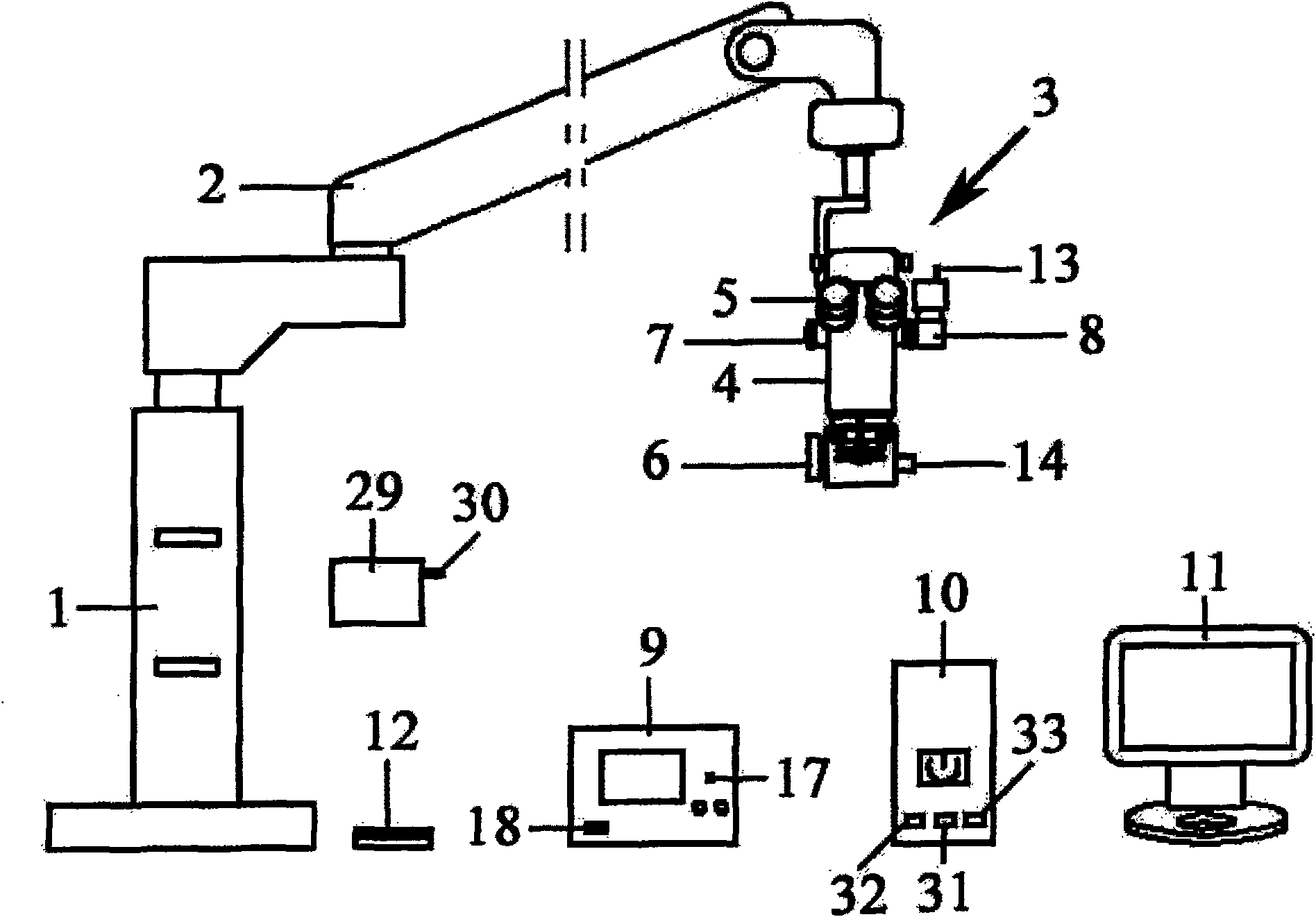
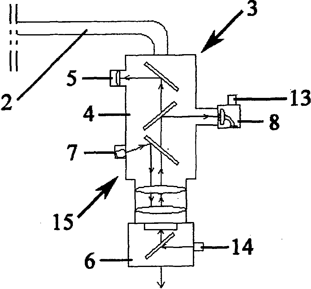
[0096] It consists of the main controller (10), display (11), CCD camera (8), laser (9), light source equipment (29), main bracket (1), cantilever (2), suspension surgical treatment head (3), display A micro observation system (4) and a microsurgery adapter (6) are composed.

[0097] The camera output terminal (13) of the CCD camera (8) is connected to the main controller (10) through a line, the laser control connection port (18) of the laser (9) is connected to the main controller (10) through a line, and the laser ( The laser output port (17) of 9) is connected to the adapter input port (14) of the microsurgery adapter (6) through an optical fiber wire, and the CCD camera (8) collects the image and spectrum of the lesion tissue and normal tissue and outputs it through the camera The terminal (13) is transmitted to the main controller (10).

[0098] The main controller (10) recognizes and processes the images and spectra of the lesion tissue and normal tissue collected by the CC...

Embodiment 2

[0108] Composed of a main controller (10), a display (11), an image capture head (25), a laser (9), a light source device (29) and an endoscope (16), a display (11) and a main controller (10) The display connection port (33) is connected to the display connection port (33), and a general display or touch screen display is used as the display (11) of the main controller (10). The display (11) instantly displays the images of the lesion tissue and normal tissue collected by the CCD camera (8) And the spectrum, while the operation parameters are displayed on the display (11).

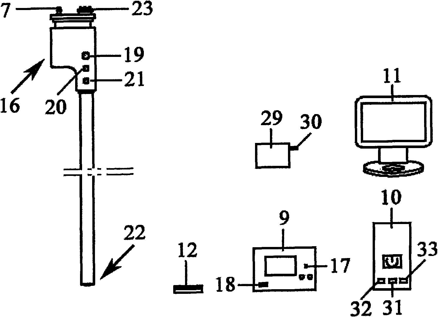
[0109] The upper part of the endoscope (16) is provided with a biopsy port (19), a stoma (20), a water hole (21), a light source interface (7) and an endoscope data port (23), a biopsy port (19), a stoma (20) ), the water hole (21) and the light source interface (7) are penetrated from the upper part of the endoscope (16) to the observation treatment end (22), and the observation treatment end (22) constitute...

Embodiment 3

[0115] After preoperative preparation, turn on the main controller (10), display (11), CCD camera (8) and light source equipment (29), adjust the cantilever (2) on the main bracket (1) and the suspension surgical treatment head (3) , The treatment end enters the operation area, and the microsurgery adapter (6) on the suspension surgical treatment head (3) is aligned with the operation site.

[0116] By hanging the image and spectrum displayed by the microscopic observation system (4) and the display (11) on the surgical treatment head (3), observe the lesion tissue and normal tissue at the surgical site, and the main controller (10) recognizes and confirms the lesion tissue and The image and spectrum of the normal tissue are used to lock the microsurgery adapter (6) to the lesion tissue site.

[0117] The laser (9) is turned on, the main controller (10) modifies the surgical parameters of the laser (9), adjusts the output power of the laser (9), and the laser (9) is ready to be tur...


PUM


Abstract

The invention provides automatic control laser operation equipment and a treatment method thereof. The equipment consists of a master controller, a monitor, a CCD camera, a laser, a light source, a master bracket, a cantilever, a hanging operation treatment head, a microscope observing system and a microscope operation adapter. The master controller recognizes and processes the image and spectrumof the focus tissue and the normal tissue collected by the CCD camera, and then gives out orders to control the work of the laser accordingly, thus avoiding operation mistakes caused by normal tissuedamage due to incorrect manipulation in laser operation; the master controller gives out orders to control the start and close of the laser and to correct the amount of laser output; a optical lens group forms a light path; and the microscope observing system, microscope inspection glass, the CCD camera and the microscope operation adapter are integrated into a whole by the optical lens group. Theinvention has simple structure, reasonable design and simple and easy operation; the normal tissue is not damaged while the focus tissue is removed in the operation; the invention is characterized inthat the equipment has a plurality of control modes; the parameters are adjustable; the application is wide. The invention can precisely control the operation extent and improve the treatment effect.

Description

Technical field [0001] The invention relates to a laser surgery medical instrument, in particular to a self-control laser surgery equipment. Background technique [0002] The use of laser microsurgery for surgery has been widely used, not only in surgery, internal medicine, ENT, and ophthalmology. In the treatment of glaucoma, laser microsurgery is the main method of treatment. Traditional filtering surgery mainly includes trabeculectomy and scleral bite surgery, but traditional filtering surgery has many postoperative complications, such as early postoperative shallow anterior chamber and no anterior chamber, ciliary artery prolapse, malignancy Glaucoma, hemorrhage, and failure to form filtering vesicles, etc.; late filtering mouth blockage, paralysis scar formation, cystic change, rupture of filtering vesicles, cataracts and infections. The existing laser microsurgery system mainly observes the changes of the biological tissues irradiated by the laser in real time through the ...

Claims


Application Information

Patent Timeline
Patent Type & Authority Patents(China)
IPC IPC(8): A61B18/20A61F9/008
Inventor 李迎新贺志江杨久敏陈谦锐徐延山薛志孝刘爱华
Owner TIANJIN MEDICAL UNIV
Why Eureka
Try Eureka