
Skull patch and its prepn process
A skull and patch technology, which is applied in the fields of cranial bone repair in neurosurgery and medical plastic surgery, can solve problems such as cerebrospinal fluid leakage, dura mater damage, and complicated surgery
- Summary
- Abstract
- Description
- Claims
- Application Information
AI Technical Summary
Problems solved by technology
Method used
Image

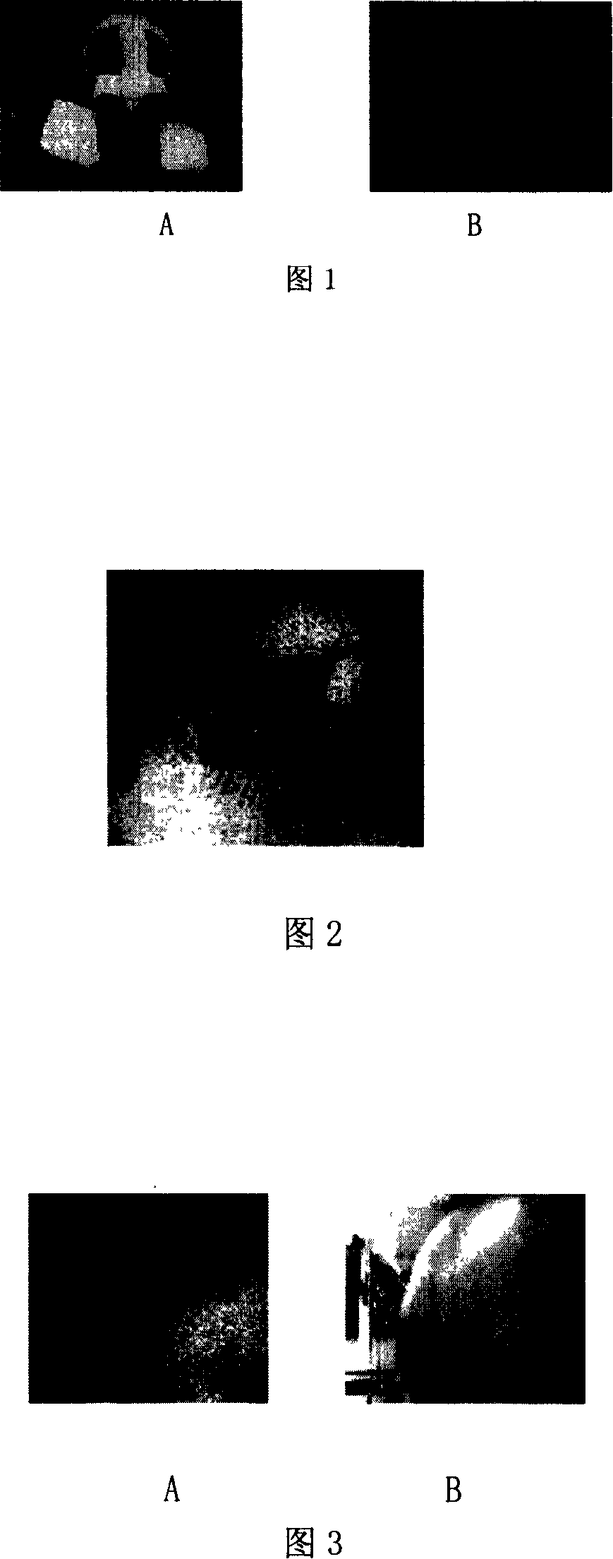
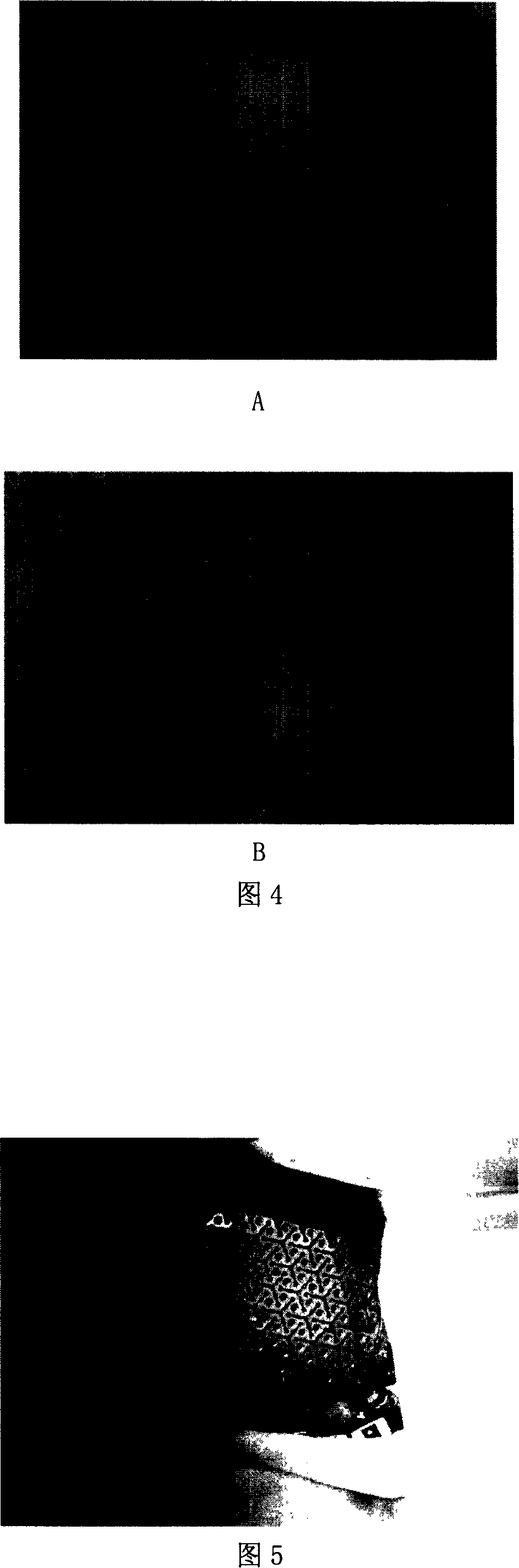
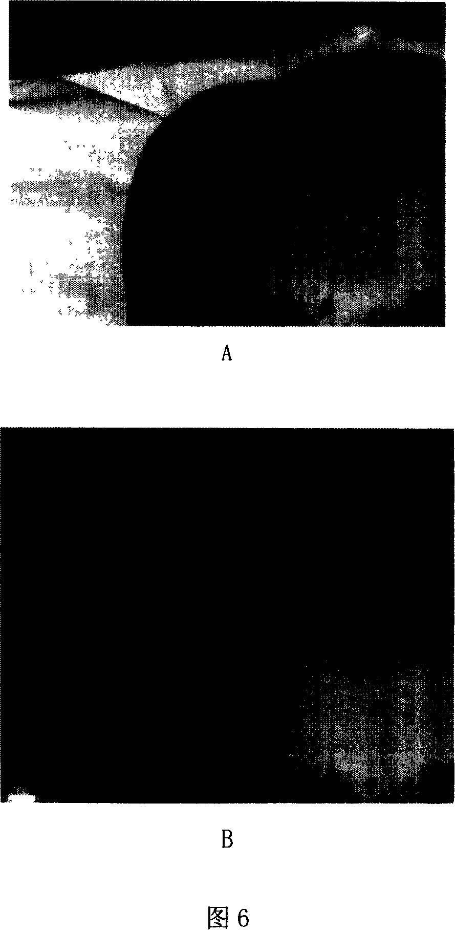
Examples
Embodiment 1
[0057] In this example, for 45 patients with a partial defect on one side of the skull and the opposite side was intact, the required titanium alloy mesh skull patch was prepared.
[0058] Use computed tomography technology to scan the image of the patient's head with a scanning distance of 0.75 mm; store the scanned image in the dicom file format on the mobile hard disk, and then transfer the data to the computer for data processing; use the image processing software minics to read Take the data, segment and extract the data information of the skull and its surrounding soft tissue (24 cases extract the skull information, 21 cases extract the skull and temporal muscle information), reconstruct the three-dimensional model of the skull and its surrounding soft tissue, and then follow the specified path, file name, stored in STL file format; use the graphics processing software magics to read the reconstructed 3D model of the skull and its surrounding soft tissues of interest; Ap...
Embodiment 2
[0060] In this example, 15 patients with bilateral skull defects were prepared to prepare the required skull patch with titanium alloy mesh.
[0061] Use computed tomography technology to scan the image of the patient's head with a scanning distance of 1.5mm; store the scanned image on a recordable CD in dicom file format, and then transfer the data to the computer for data processing; use the image processing software minics Read the data, segment and extract the data information of the skull and its surrounding soft tissues (9 cases extract the skull information, 6 cases extract the skull and temporal muscle information), reconstruct the three-dimensional model of the skull and its surrounding soft tissues, and then follow the specified path, File name, stored in STL file format; use the graphics processing software magics to read the reconstructed 3D model of the skull and its surrounding soft tissues of interest; use the graphics processing software magics to read the shape...
Embodiment 3
[0063] In this example, for 10 patients with only one side of the skull partially defective and the opposite side intact, the required silicone rubber polyester mesh skull patch was prepared.
[0064]Use computed tomography technology to scan the image of the patient's head with a scanning distance of 0.75 mm; store the scanned image in the dicom file format on the mobile hard disk, and then transfer the data to the computer for data processing; use the image processing software minics to read Take data, segment and extract the data information of the skull and its surrounding soft tissues (including 7 cases of skull information extraction, of which 3 patients with unilateral temporal skull injury, extraction of skull and temporal muscle information), reconstruction of the skull and its surrounding soft tissues of interest Then store it in STL file format according to the specified path and file name; use the graphics processing software magics to read the reconstructed 3D prot...
PUM
 Abstract
Description
Claims
Application Information