Data acquisition system for nuclein and fluorescent dual module integral small animal molecules imaging

A dual-mode, molecular imaging technology, used in medical science, sensors, inoculation and ovulation diagnosis, etc., can solve problems such as difficult coupling between crystals and photomultiplier tubes, expensive special photomultiplier tubes, and inability to perform tomographic imaging of small animals. Achieve low cost, reduce radiation damage, and achieve simple results

Inactive Publication Date: 2007-10-24
TSINGHUA UNIV
View PDF0 Cites 38 Cited by
  • Summary
  • Abstract
  • Description
  • Claims
  • Application Information

AI Technical Summary

Problems solved by technology

Although this solution can realize dual-mode detection, this system can only realize the self-luminescence measurement of animals, without the function of fluorescence imaging
Moreover, it is only planar imaging, and cannot perform tomographic imaging of small animals as a whole
In addition, this system requires a special photomultiplier tube, which is expensive, and the coupling between the crystal and the photomultiplier tube is also difficult, and the electronic system required for the separation of the two signals is also quite complicated.

Method used


Image

  • Data acquisition system for nuclein and fluorescent dual module integral small animal molecules imaging
  • Data acquisition system for nuclein and fluorescent dual module integral small animal molecules imaging
  • Data acquisition system for nuclein and fluorescent dual module integral small animal molecules imaging

Examples


Embodiment Construction

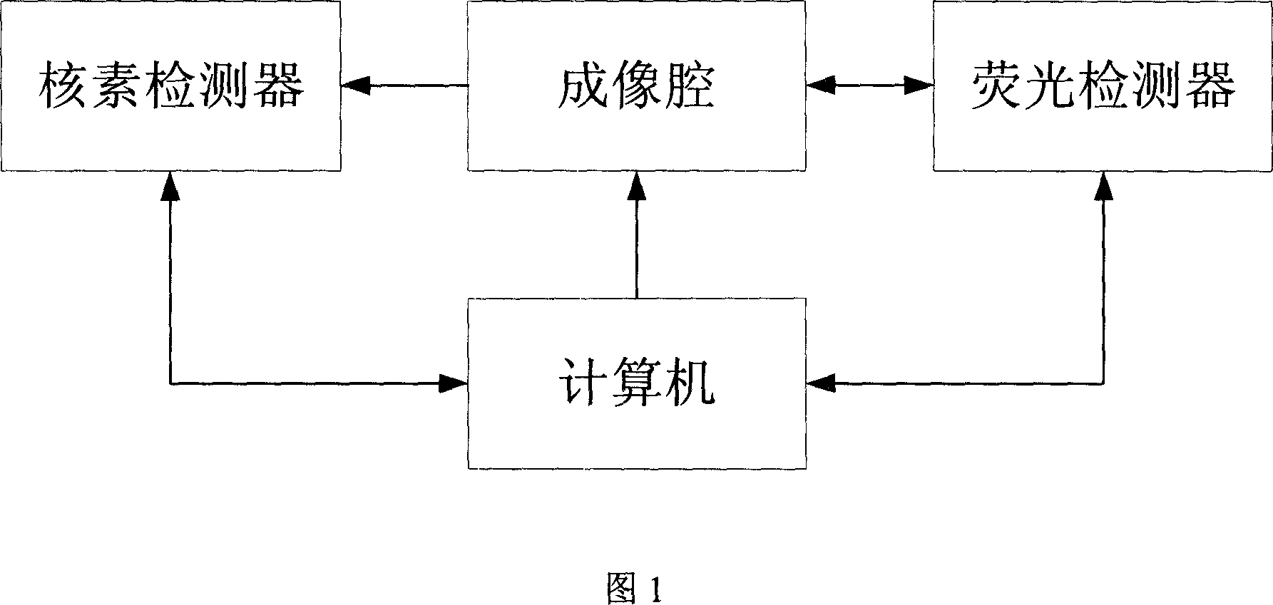
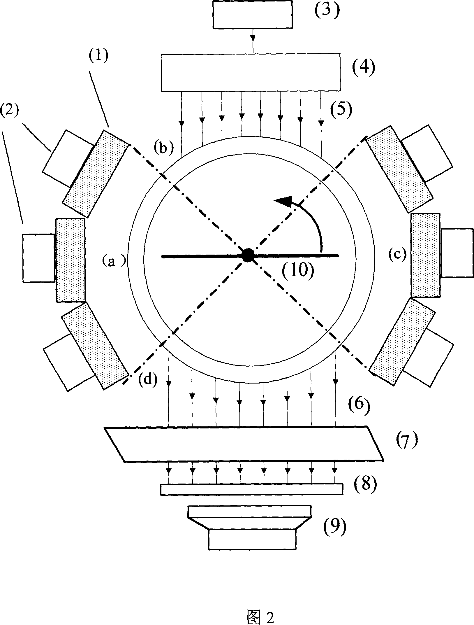
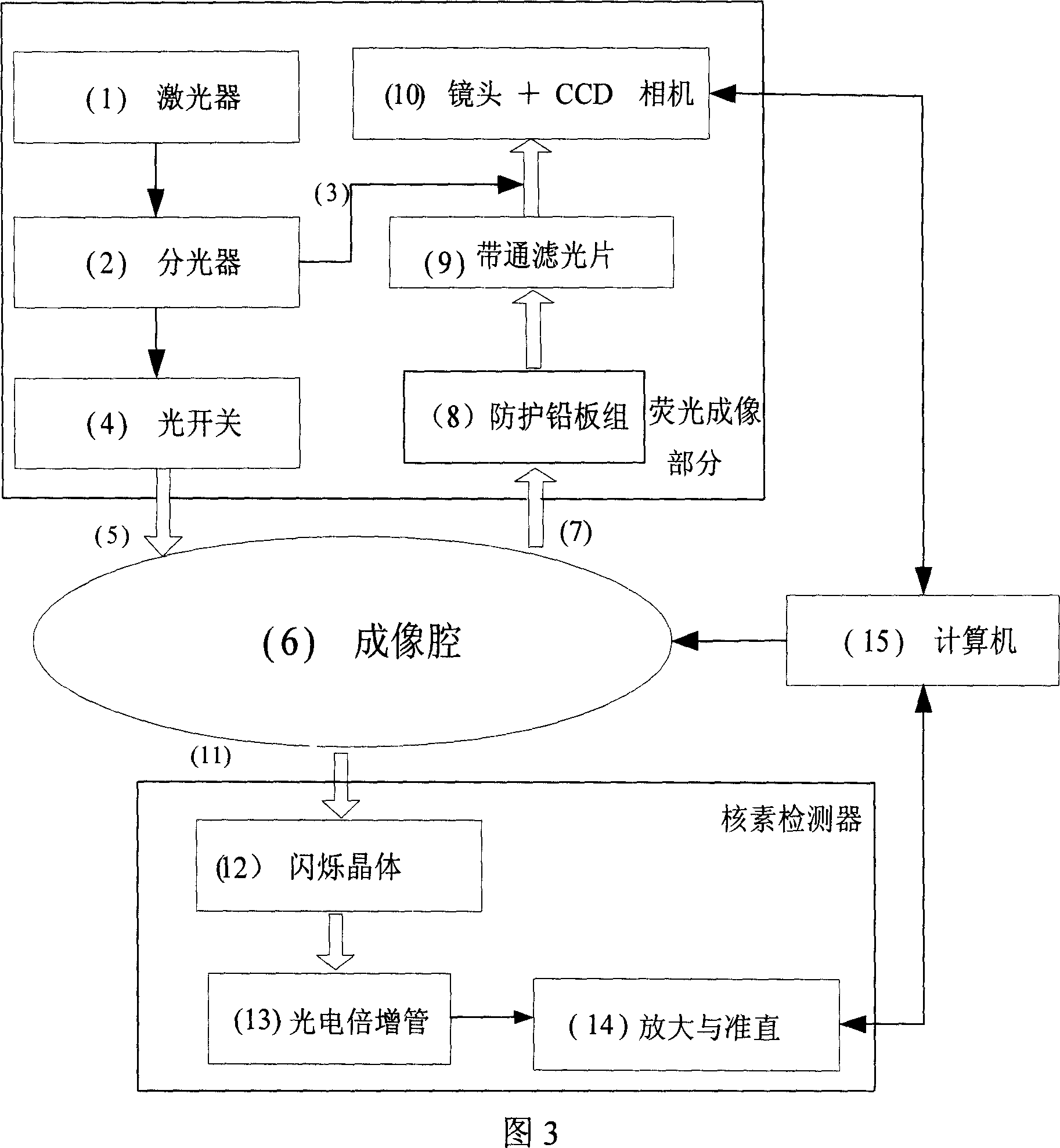
[0024] The dual-mode imaging system described in the present invention mainly includes the following four basic steps: 1) Marking preparation stage. Inject specific nuclides and fluorescent markers into living small animals to label cells or tissues. 2) The tested animal is fixed. After a certain reaction and aggregation time, the small animal is anesthetized and suspended on the bracket in the imaging cavity, and its limbs need to be fixed at the same time. The cavity can be filled with optical medium liquid (such as fat emulsion solution) as required. 3) Data collection stage. The bracket suspended by the animal is controlled by the stepping motor at the bottom of the imaging chamber to rotate around the vertical direction at a low speed. At this time, the nuclide detector and the fluorescence detector located in an orthogonal distribution around the imaging chamber collect signals respectively, and the raw data It is sent to the computer through the control interface car...


PUM


Abstract

The invention belongs to the field of application of near-infrared laser, nuclear irradiation, electron and image rebuild in systematic biology and medicine. It is characterized in that high-specificity nuclear species and fluorescent probe are implanted in live animal to realize double mark of muscle deep layer or internal organ tumor cell. The emission signal of nuclear species is checked by sensitive electron-multiplier phototube in array copulation position of scintillation crystal; feeble fluorescent signal generated by visible light or infrared agitation is checked by high quantum yield CCD camera. The two checking systems are orthogonally arranged in the same surface, the live nuclear species and fluorescent signal can be checked simultaneously through rotary image cavity, and double module three dimensional tomography image of small animal can be rebuilt with software algorism. The invention is characterized by multi-image forming modules, abundant information and simple operation.

Description

technical field [0001] The data acquisition system for molecular imaging of small animals with nuclide and fluorescence dual-mode integration belongs to the comprehensive technical field of laser, nuclear radiation, electron and image reconstruction in the application of systems biology and medicine. Background technique [0002] The invention belongs to one of molecular image application technologies. After medical imaging technology experienced structural imaging and functional imaging, since the 1990s, with the development of biological and genetic technology and the improvement of imaging technology, new molecular imaging technology has emerged. It can realize non-invasive, dynamic and real-time in vivo imaging of physiological or pathological processes inside organisms at the gene, cell and molecular levels, so as to provide insights into disease-related gene function localization, cell growth and mutation process mechanism, new drug development, tumor Treatment and ot...

Claims


Application Information

Patent Timeline
Patent Type & Authority Applications(China)
IPC IPC(8): A61B10/00A61B19/00A61B5/00A61B90/00
Inventor 白净黄国亮单保慈宋小磊胡刚张永红奉华成尚尚汪待发
Owner TSINGHUA UNIV
Why Eureka
Try Eureka