Method and material for simultaneously detecting mycobacterium tuberculosis compound and identifying its medicament resistance

A technology of mycobacterium tuberculosis and mycobacteria, applied in the fields of molecular biology, medicine, and microbiology, can solve the problems of expensive, difficult to prepare probe sets, and inability to detect the sensitivity of pathogens, achieving short-term and low-cost Effect

Inactive Publication Date: 2008-07-02
RUSN ACAD V A ENGERHARDT MOLECULAR BIOLOGY INST
View PDF0 Cites 10 Cited by
  • Summary
  • Abstract
  • Description
  • Claims
  • Application Information

AI Technical Summary

Problems solved by technology

[0072] - the application of the ligation-based method (XV) is limited due to the difficulty in preparing probe sets for a large number of detection targets and the use of expensive enzymes (thermostable DNA-ligases);
[0073] - Real-time PCR (XVI) only reveals the presence of the most widespread mutations in the tested genomic fragments, but cannot precisely identify nucleotide substitutions, and the method is expensive relative to routine analysis;
[0074] - Fluorescence resonance energy transfer (XVIII) has two significant disadvantages: 1) it only allows the detection of a limited number of mutations, 2) it does not allow the distinction of functionally important mutations from those that are not manifested phenotypically
[0075] -The method of PCR and ligation on oligonucleotide microchips (XIX) needs further improvement and is somewhat complicated for practical laboratory application
[0076] All the above-mentioned methods based on amplification of IS-factor (XX), heat shock protein (XX), detection of M. tuberculosis complex based on ribosomal spacer gene detection (XXI), and automated PCR (XXII), The methods of ligase chain reaction (XXIII) and real-time PCR (XXIV) allow the detection of the presence of the tuberculosis pathogen in the tested sample, but not the susceptibility of the pathogen to antimycobacterial drugs

Method used


Image

  • Method and material for simultaneously detecting mycobacterium tuberculosis compound and identifying its medicament resistance
  • Method and material for simultaneously detecting mycobacterium tuberculosis compound and identifying its medicament resistance
  • Method and material for simultaneously detecting mycobacterium tuberculosis compound and identifying its medicament resistance

Examples


Embodiment 1

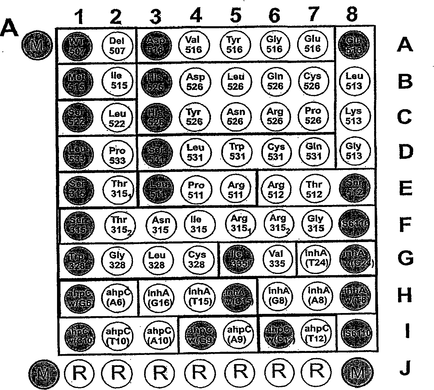
[0123] Example 1. Biochip for detection of Mycobacterium tuberculosis complex and evaluation of strain sensitivity to rifampicin and isoniazid

[0124]1. The oligonucleotides for immobilization on the biochip and the primers for amplification are synthesized on the automatic synthesizer 394DNA / RNA synthesizer (Applied Biosystems, USA), and contain the oligonucleotides for further immobilization on the gel respectively. The free amino 3′-amino-modifier C7 CPG 500 (Glen Research, USA) on the fluorochrome or the spacer for the 5′-amino-modifier C6 (Glen Research, USA) for attaching the fluorescent dye. Attachment of indodicarbocyanine fluorescent dye ("Biochip-IMB", Russia) was performed according to the manufacturer's recommendations. Biochips were produced according to methods described earlier (Rubina AY, Pan'kov SV, Dementieva EI et al., Hydrogel drop microchips with immobilized DNA: properties and methods for large-scale production. AnalBiochem 2004; 325: 92-106). The bioch...

Embodiment 2

[0143] Example 2. Processing of clinical samples

[0144] 1. Mix clinical samples (sputum, exudates, eluate, bronchoalveolar lavage) with a freshly prepared 0.5% solution of N-acetyl-L-cysteine ​​(NALC) in 2% NaOH Mix in a 1:1 (v / v) ratio. The samples were stirred vigorously with a Vortex and maintained at room temperature for 20 minutes. Phosphate buffered saline pH 6.8 was added to the sample at a ratio of 1:5 (v / v), and the mixture was centrifuged at 3,000 rpm for 30 minutes. When using cerebrospinal fluid, perform a preliminary centrifugation at 10,000 rpm for 10 minutes. For blood analysis, the lymphocyte fraction was initially isolated according to the usual method using Ficoll. Subsequent processing was the same for all samples.

[0145] 2. The precipitated pad was suspended with 1.5 mL of TE buffer (10 mM Tris-HCl, 1 mM EDTA) (pH 8.0), and then centrifuged at 3,000 rpm for 30 minutes. Repeat the washing process several times.

[0146] 3. Add 30 μl of TE buffer (p...

Embodiment 3

[0148] Example 3. Amplification of fragments of IS6110 movable factor, rpoB, katG, inhA, ahpC genes; preparation of single-stranded fluorescently labeled fragments by multiplex PCR

[0149] In the first step, multiplex amplification of fragments of the rpoB (212 b.p.), katG (166 b.p.), inhA (133 b.p.), ahpC (126 b.p.) genes and the IS6110 mobile factor (309 b.p.) was performed.

[0150] 3 μl of the sample obtained in Example 2, paragraph 4, were added to 25 μl of the PCR-mix.

[0151] Composition of the PCR-Mix:

[0152] 1X PCR-buffer: 10 mM KCl, 10 mM Tris-HCl (pH 8.3) (Sileks, Russia);

[0153] 1.5mM MgCl 2 ;

[0154] dATP, dCTP, dGTP, and dUTP (Sileks) are each 200 μM;

[0155] A mixture of primers (sequences listed in Table 2) at the following concentrations: p105f, p293r, katG_f, katG_r1, IS_f, IS_r1; InhA_f, InhA_r1, ahpc_f, ahpC_r1 - 100 nM;

[0156] 5U thermostable Taq DNA polymerase (sileks);

[0157] • 0.5 U uracil-DNA-glycosylase (Sileks).

[0158] Amplify on...


PUM

PropertyMeasurementUnit
diameteraaaaaaaaaa

Abstract

The present invention relates to molecular biology, microbiology and medicine, and provides a method for detecting nodule offshoot bacilli complex body in clinic samples and evaluating the sensitivity of strain to rifampicin as well as isoniazid in distinguish biochip. The method obtains fluorescence DNA segments based on two-step multiple PCR, then to hybridize said segments on a micro-array containing differential distinguished oligonucleotide group. To determine the resistance of concretion offshoot bacilli to rifampicin and isoniazid by means of evaluating point nucleotide replacement in microbiology DNA. The present invention allows analyze directly clinic sample to evaluate mass mutation at one time, so as to reduce analysis cost and implement time. The present invention also relates to primer group, biochip and oligonucleotide probe group for implementing said method.

Description

field of invention [0001] The present invention relates to molecular biology, microbiology, and medicine, and provides detection of Mycobacterium tuberculosis (Mycobacterium tuberculosis) complex (Mycobacterium tuberculosis, Mycobacterium bovis (M. ), Mycobacterium bovis BCG, Mycobacterium africanum (M.africanum), and Mycobacterium microti (M.microti)) and simultaneously evaluate the sensitivity of strains to rifampicin and isoniazid. Background of the invention [0002] The following methods are commonly used to detect mutations that confer drug resistance: [0003] I. Single Nucleotide Polymorphism (SNP); [0004] Dubiley S. Kirillov E and A. Mirzabekov. 1999. Polymorphismanalysis and gene detection by minisequencing on an array of gel-immobilized primers. Nucleic Acids Research, Vol.27, No.18(e19). [0005] Marth GT, Korf I, Yandell MD et al., 1999. A general approach to single-nucleotide polymorphism discovery. Nat Genet. Dec;23(4):452-456. [0006] II. Allele-specifi...

Claims


Application Information

Patent Timeline
Patent Type & Authority Applications(China)
IPC IPC(8): C12Q1/68C12Q1/04
CPCC12Q2600/16G01N2333/35C12Q1/689
Inventor A·S·加塞达特勒夫A·Y·索博勒夫D·A·格亚杜诺夫S·A·拉帕V·M·米卡伊洛维奇A·D·米尔加贝科夫
Owner RUSN ACAD V A ENGERHARDT MOLECULAR BIOLOGY INST
Why Eureka
Try Eureka