
RNA quantitative determination method utilizing S1 enzyme cutting single-chain nucleic acid characteristic
A quantitative detection method and cutting sheet technology, applied in the field of bioengineering, can solve the problems of cumbersome steps in sandwich hybridization technology, lack of specificity of detection, increase of detection cost, etc., and achieve stable results, simple method, and easy operation
Patent Information
- Authority / Receiving Office
- CN · China
- Current Assignee / Owner
- Publication Date
- 2008-09-10
- Estimated Expiration
- Not applicable · inactive patent
Smart Images
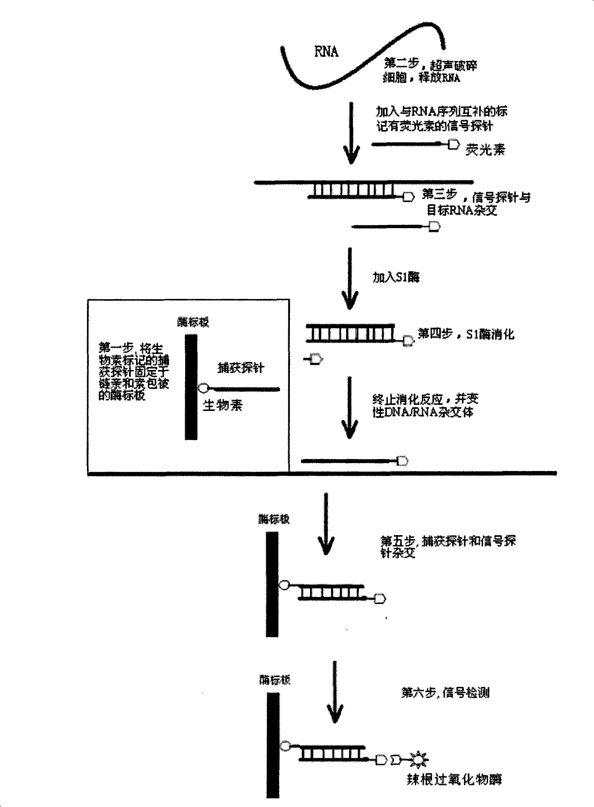
Figure 1 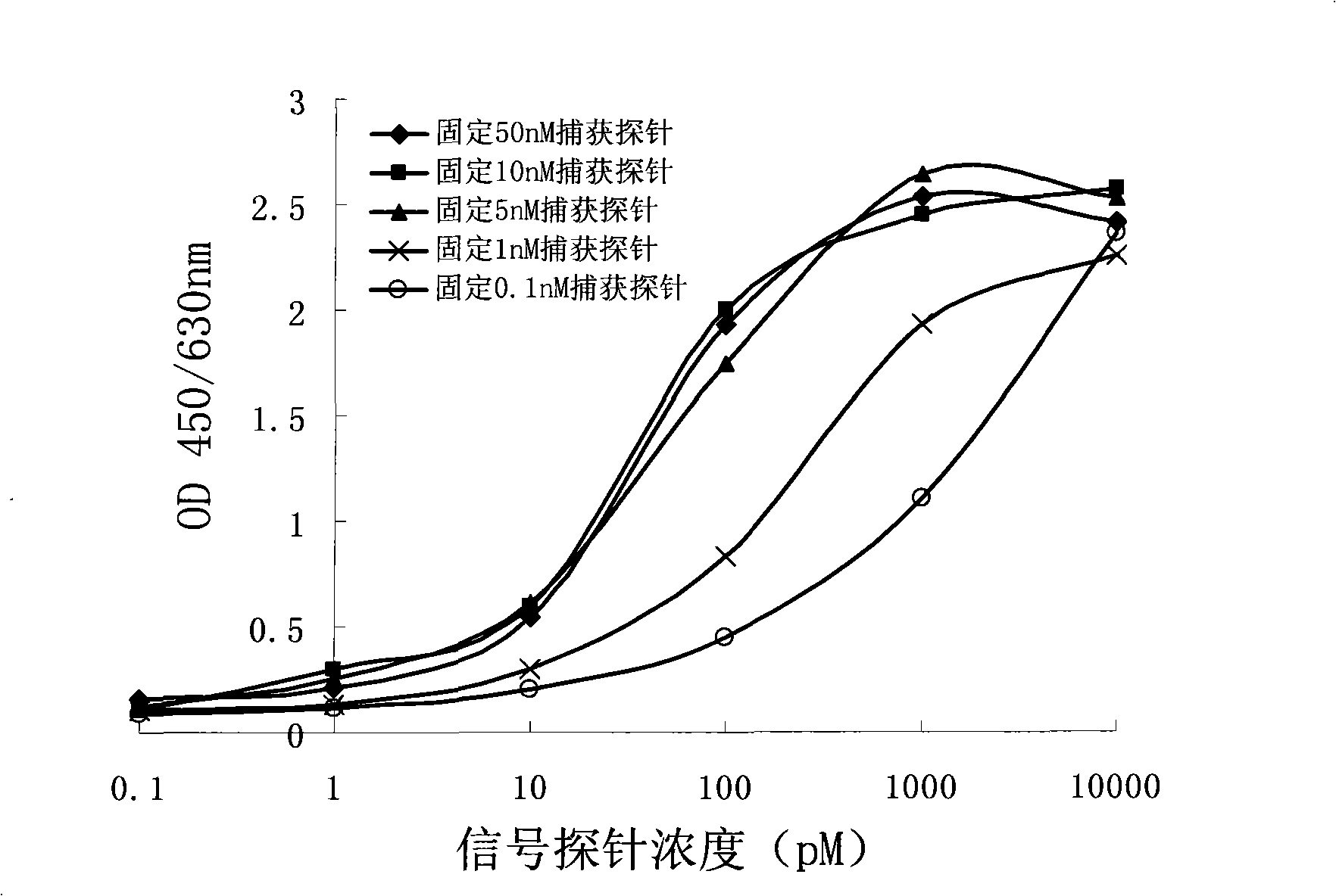
Figure 2 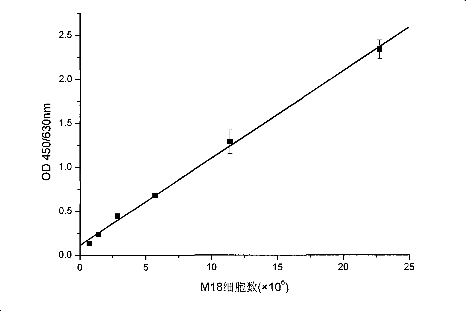
Figure 3
Abstract
Description
technical field
[0001] The invention relates to a detection method, in particular to an RNA quantitative detection method using S1 enzyme to cut single-stranded nucleic acid characteristics, and belongs to the technical field of bioengineering. Background technique
[0002] More and more attention has been paid to the development of a rapid, sensitive and reliable genetic detection method. Traditional Northern hybridization and slot hybridization take too long and the operation is very complicated. Although the gene chip technology developed in recent years can detect gene expression with high throughput, it is a costly method. Fluorescent quantitative PCR technology has high detection sensitivity, it needs to purify very high quality RNA, otherwise it will inhibit the subsequent reverse transcription and PCR reactions. The detection of sandwich hybridization technology is fast, even without nucleic acid purification, which is lacking in the specificity of detection. [0...
Examples
Embodiment 1
[0028] Effects of different concentrations of capture probes on the detection signal:
[0029] 1.1. Design signal probes and capture probes for Pseudomonas fluorescens:
[0030] Search the sequence of AY394845 in NCBI, and get AY394845, Pseudomonas sp.M18 16Sribosomal RNA gene, partial sequence. Input the obtained sequence into the software primerprimier 5 for probe search, set the probe length to 40+-3bp (other parameters default to ), select probes with GC content of 40-60% according to rating from high to low in the obtained probes, and obtain 39 bases of probes against M1816s RNA:
[0031] Capture probe:
[0032] 5'-(biotin)-AGGGTGCAAGCGTTAATCGGAATTACTGGGCGTAAAGCG
[0033] Signal probe:
[0034] 5’-(FAM)-CGCTTTACGCCCAGTAATTCCGATTAACGCTTGCACCCT
[0035] 1.2. Capture probe immobilization:
[0036] Dilute with phosphate buffer solution to obtain capture probes with a concentration of 0.1nM, 1nM, 5nM, 10nM and 50nM, respectively take 100μl and add them to the enzyme plate...
Embodiment 2
[0046] The detection limit and linear range of the 16s RNA of Pseudomonas fluorescens M18 (the Pseudomonas fluorescens strain M18 used has been designated by the Chinese Patent Office as a depository unit: Beijing, China Microorganism Culture Collection Management Committee General Microbiology Center, deposit number CGMCC NO.0462, 2000.6.27):
[0047] 2.1. Design signal probes and capture probes for Pseudomonas fluorescens:
[0048] Search the sequence of AY394845 in NCBI, and get AY394845, Pseudomonas sp.M18 16Sribosomal RNA gene, partial sequence. Input the obtained sequence into the software primerprimier 5 for probe search, set the probe length to 40+-3bp (other parameters default to ), select probes with GC content of 40-60% according to rating from high to low in the obtained probes, and obtain 39 bases of probes against M18 16s RNA:
[0049] Capture probe:
[0050] 5'-(biotin)-AGGGTGCAAGCGTTAATCGGAATTACTGGGCGTAAAGCG
[0051] Signal probe:
[0052] 5'-(FAM)-CGCTTTAC...
Embodiment 3
[0070] Determination of 16s RNA content in different growth stages of Pseudomonas fluorescens M18:
[0071] 3.1. Bacterial culture and plate count:
[0072] Inoculate the plate-activated M18 strain into 100ml of KB liquid medium, culture it on a shaker at 28°C and 160rpm, and use the plate counting method to detect the bacterial concentration at 12 hours, 24 hours, 36 hours and 60 hours, and liquid nitrogen quick-freezing Save the bacteria. The bacterial concentration measured by the plate counting method was 0.97×10 10 CFU / ml, 2.5×10 10 CFU / ml, 7.4×10 10 CFU / ml and 9.5×10 10 CFU / ml.
[0073] 3.2. Capture probe immobilization:
[0074] Add 100 μl of 5 nM capture probe diluted with phosphate buffer to a streptavidin-coated microtiter plate, bind at 37°C and 200 rpm for 2 hours, wash with phosphate buffer 3 times, and store at 4°C for use.
[0075] 3.3. Bacterial fragmentation:
[0076] Take a portion of the M18 cells (centrifuged from 1ml of the bacteria liquid) that we...