Method and apparatus realizing quasi confocal fluorescent microscopic with dynamic speckle illumination

A fluorescence microscope and speckle technology, applied in the field of confocal fluorescence microscopy, can solve problems such as unsuitable sample imaging, and achieve the effects of easy operation and promotion, simple device structure, and convenient later data processing.

Inactive Publication Date: 2008-11-12
SHENZHEN UNIV
View PDF0 Cites 32 Cited by
  • Summary
  • Abstract
  • Description
  • Claims
  • Application Information

AI Technical Summary

Problems solved by technology

This microscopic imaging technique has tomographic imaging capabilities similar to traditional confocal microscopic imaging techniques, but it is not suitable for imaging samples with a certain thickness

Method used


Image

  • Method and apparatus realizing quasi confocal fluorescent microscopic with dynamic speckle illumination
  • Method and apparatus realizing quasi confocal fluorescent microscopic with dynamic speckle illumination
  • Method and apparatus realizing quasi confocal fluorescent microscopic with dynamic speckle illumination

Examples


Embodiment Construction

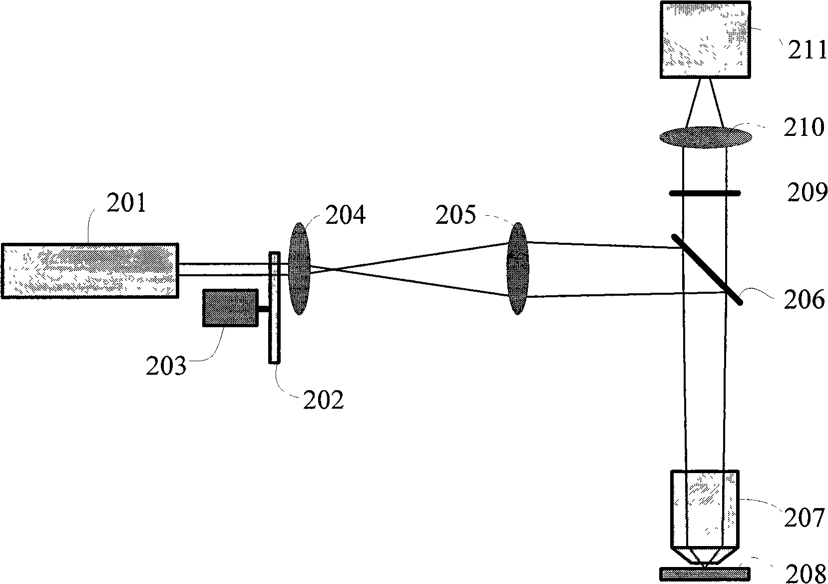
[0046] The specific embodiment of the present invention is as figure 2 shown. An argon ion laser 201 with a wavelength of 488 nm is used as a light source, and the average output power is 20 mW. The laser light emitted by 201 passes through a glass substrate scatterer 202 with a specific particle size to generate a speckle pattern. The 202 is mounted on a stepping motor 203, and the 203 is controlled by a computer to step and rotate by about 0.5 degrees to generate a dynamic speckle pattern. The generated speckle illumination excitation beam is coupled to a traditional wide-field fluorescence microscope after being beam expanded and shaped, coupled with the relay optical system 204 and 205, and the excitation light is reflected into the microscope after passing through the dichroic beam splitter 206. The objective lens 207 is focused on the sample 208 to form dynamic speckle illumination. The fluorescent signal emitted by the sample passes through the dichroic mirror 206 ,...


PUM


Abstract

The present invention relates to a novel method and a novel device, incorporating dynamic speckle lighting with a conventional wide field fluorescence microscope organically to implement approximately confocal fluorescence microscopy (i.e., quasi-confocal fluorescence microscopy). The present invention employs an argon ion laser as the light source; the exciting light passes through a scattering object, a relay light path system for expanding and shaping, and then is coupled to the fluorescence microscope and focused to the sample. The stepping and revolution of the scattering object is controlled with a computer to produce a dynamic speckle pattern on the sample. The received fluorescent images are processed to obtain high-resolution spatial chromatographic images under no need to scan condition. The method can be used to obtain high-resolution three-dimensional chromatographic and microscopic image information of biological tissue samples in a non-intrusive manner. The device has simple structure, high cost-performance ratio, and is favorable for post data processing, easy to operate and apply; therefore, the device and method have a wide market application prospect and great significance for clinical diagnosis and life science research.

Description

technical field [0001] The invention is a new method and a new device for approximately realizing confocal fluorescence microscopy (that is, quasi-confocal fluorescence microscopy) by utilizing dynamic speckle illumination. This method can obtain high temporal and spatial resolution three-dimensional tomographic microscopic image information of biological tissue samples in a non-invasive manner. The device has a simple structure, high cost performance, convenient post-data processing, easy operation and promotion, and has huge application prospects and The market is of great significance to clinical disease diagnosis and life science research. Background technique [0002] In the field of life science research and clinical diagnosis, laser scanning confocal microscopy [1] has become a microscopic technique that can effectively obtain high spatial resolution three-dimensional tomographic image information of biological tissue samples. Compared with traditional optical micros...

Claims


Application Information

Patent Timeline
Patent Type & Authority Applications(China)
IPC IPC(8): G01N21/64G01N21/47
Inventor 屈军乐尹君赵羚伶王磊牛憨笨
Owner SHENZHEN UNIV
Why Eureka
Try Eureka