Immune colloidal gold test paper strip for detecting yapamicin relict and preparation thereof

A technology of colloidal gold test paper and kanamycin, which is applied in the field of immunochemical rapid detection of veterinary drug residues, can solve the problems of unfavorable promotion and use, complicated operation process, and cumbersome measurement methods, and achieve short detection time, low temperature requirements, The effect of easy operation

Inactive Publication Date: 2008-12-03
CHINESE ACAD OF INSPECTION & QUARANTINE
0 Cites 36 Cited by

AI-Extracted Technical Summary

Problems solved by technology

Chromatography or chromatography-mass spectrometry has high sensitivity and reliable results, but the determination method is cumbersome, time-consuming, and costly, and operators need professional training, so it is difficult to popularize
ELISA is a competitive immunoassay method using enzyme markers, which has been widely used as a rapid screening method for qualitative or semi-q...

Method used

Kanamycin, gentamicin, neomycin, streptomycin, chloramphenicol, cephalexin, sulfadimethoxine, enrofloxacin are mixed with the sample that concentration is 10ng/mL, will The drop is tested on the sample pad or the sample hole. Result of the test (seeing table 1) shows that the kanamycin sample detection line has no color, and a red band appears on the quality control line simultaneously, while gentamicin, neomycin...

Abstract

The invention relates to a test strip used for detecting immune colloidal gold remaining in kanamycin. The test strip comprises a sample pad, a combination pad, a cellulose nitrate membrane, a water absorbing pad and a PVC back lining, wherein, the sample pad, the combination pad, the cellulose nitrate membrane and the water absorbing pad are continuously stuck on the PVC back lining in sequence,lapping is arranged between the combination pad and the cellulose nitrate membrane, and lapping is arranged between the cellulose nitrate membrane and the water absorbing pad; an anti-kanamycin monoclonal antibody-colloidal gold label is coated on the combination pad, and a detection line formed by a kanamycin-carrier protein (bovine serum albumin) coupling compound and a quality controlling lineformed by a rabbit anti mouse are coated on the cellulose nitrate membrane. The test strip of the invention has the advantages that the specificity is strong, the sensitivity is high, the detection time is short (10 minutes), the test strip can be operated on site, the detection cost is low, the operation is simple, and the test strip is not required to be operated by professionals.

Application Domain

Technology Topic

Image

  • Immune colloidal gold test paper strip for detecting yapamicin relict and preparation thereof
  • Immune colloidal gold test paper strip for detecting yapamicin relict and preparation thereof
  • Immune colloidal gold test paper strip for detecting yapamicin relict and preparation thereof

Examples

  • Experimental program(4)

Example Embodiment

[0030] Example 1: The structure of the immune colloidal gold test strip of the present invention
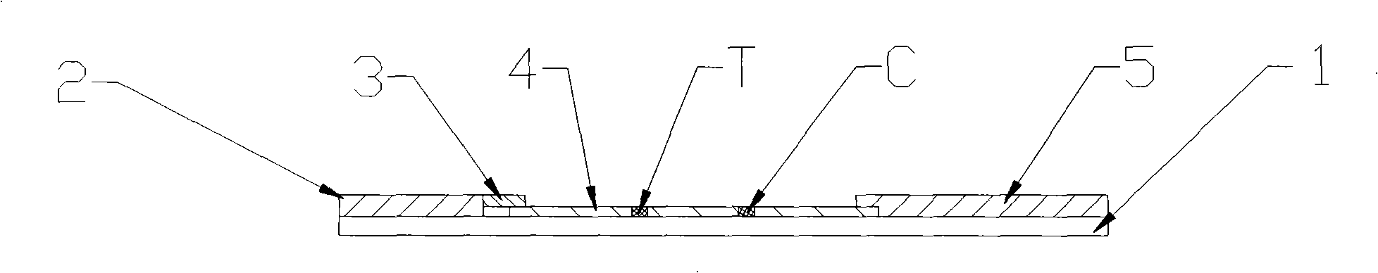
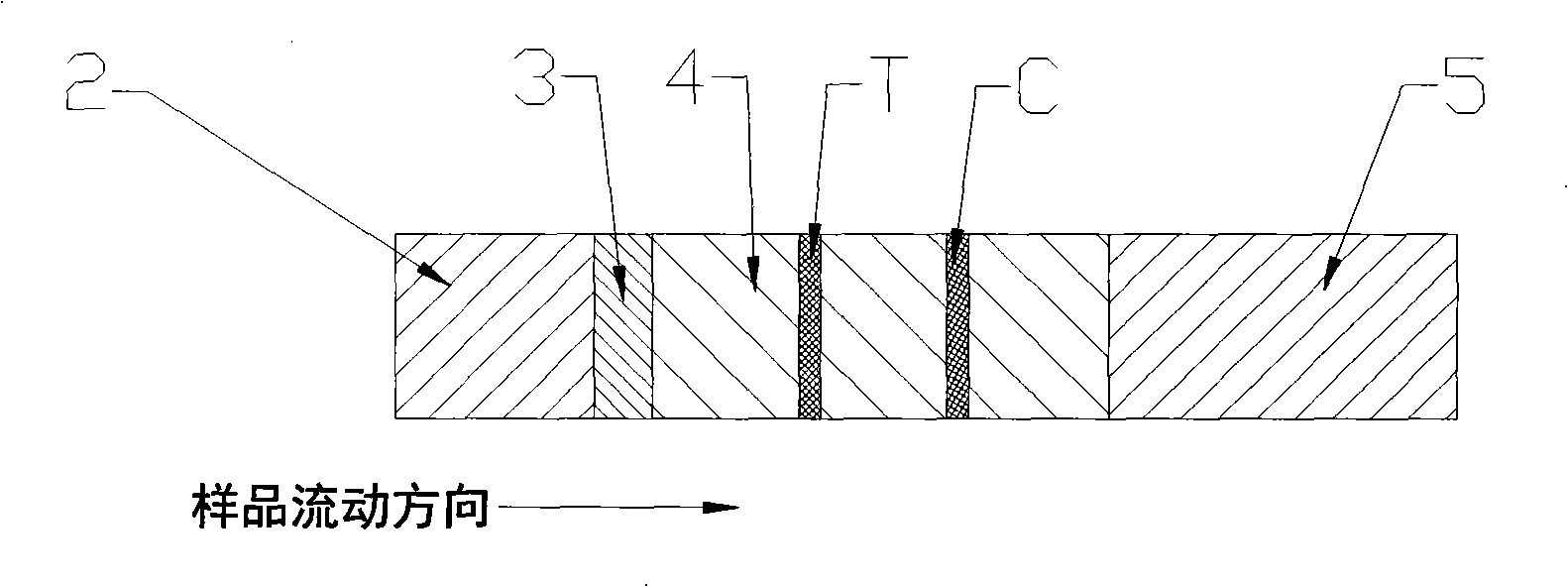
[0031] Such as figure 1 with figure 2 As shown, an immune colloidal gold test strip for detecting kanamycin residues, including sample pad 2, binding pad 3, nitrocellulose membrane 4, absorbent pad 5 and PVC backing 1, in the PVC backing 1. The sample pad 2, the binding pad 3, the nitrocellulose membrane 4 and the absorbent pad 5 are successively adhered to the upper part. There is an overlap between the binding pad 3 and the nitrocellulose membrane 4, and between the nitrocellulose membrane 4 and the absorbent pad 5 Overlap; the binding pad 3 is coated with anti-kanamycin monoclonal antibody-colloidal gold marker, and the nitrocellulose membrane 4 is respectively coated with kanamycin-carrier protein (bovine serum albumin) coupling The detection line T formed by the substance and the quality control line C formed by the rabbit anti-mouse IgG. Finally cut into 3mm wide strips and vacuum package.
[0032] When in use, the sample can be dripped onto the sample pad after a certain treatment. Special sample holes can be made on the sample pad.
[0033] The invention selects kanamycin-carrier protein conjugate as the detection line, the anti-kanamycin specific monoclonal antibody is a colloidal gold-labeled antibody, and the competition method is used to detect whether the sample to be tested contains kanamycin. The kanamycin in the sample to be tested and the kanamycin-carrier protein conjugate coated on the nitrocellulose membrane compete with the anti-kanamycin monoclonal antibody-colloidal gold marker, which appears at the detection line Red bands with different shades or no bands appear, and red bands appear at the quality control line. If the color of the test strip of the sample to be tested is significantly lighter than the color of the test strip of the negative sample, and a red band appears on the quality control line, it is judged as a positive sample, that is, the concentration of kanamycin in the sample to be tested is 1 ~10ng/mL; if the test strip of the sample to be tested has no color on the test line, and a red band appears on the quality control line, it is also judged as a positive sample, that is, the concentration of kanamycin in the sample to be tested is higher than 10ng/ mL; If the color of the test strip of the sample to be tested is equal to or darker than the color of the negative sample test strip, and a red band appears on the quality control line, it is judged as a negative sample, that is, the concentration of kanamycin is less than 1ng/ mL; if there is no red band on the quality control line, the test strip is invalid (such as image 3 Shown).

Example Embodiment

[0034] Example 2 (Preparation Example)
[0035] Preparation method of immune colloidal gold test strip for detecting kanamycin
[0036] 1. Preparation of kanamycin-carrier protein conjugate
[0037] Synthesis of immunogen: weigh 45 mg kanamycin sulfate and 10 mg hemocyanin (KLH), dissolve in 1.5 mL PBS, and stir on a magnetic stirrer. Dissolve 300 mg of EDC·HCl in 1 mL of PBS, adjust the pH to 7.4, and add it dropwise to the above mixed solution, stir at room temperature for 1 hour, and then stand at 4°C for 3 days. It was dialyzed against PBS for 3 days, the dialysate was changed 2 to 3 times a day, and further purified by a Sephadex G-25 column, packed in aliquots, and stored at -20°C.
[0038] Synthesis of the coating agent: Weigh 45 mg of kanamycin sulfate and 10 mg of BSA in 1.5 mL of PBS, and stir on a magnetic stirrer. Dissolve 300 mg of EDC·HCl in 1 mL of PBS, then add it dropwise to the above mixed solution, and stir and react at room temperature for 3 hours. It was dialyzed with PBS for 3 days, the dialysate was changed 2 to 3 times a day, and further purified by Sephadex G-25 column, aliquoted, and stored at -20°C.
[0039] 2. Preparation of anti-kanamycin monoclonal antibody
[0040]The kanamycin-hemocyanin (KLH) conjugate prepared by the applicant was used to immunize 4 8-week-old BAL/C mice (purchased from the Experimental Animal Center of the Academy of Military Sciences). 100μg solution of KLH conjugate was emulsified with an equal volume of Freund's complete adjuvant (purchased from Sigma), injected into the abdominal cavity of mice, 15 days after immunization, replaced with Freund's incomplete adjuvant for emulsification (Purchased from Sigma company) booster immunization, once every 15 days, a total of 3 times, 10 days after each immunization, blood was collected from the posterior orbital vein of the mouse to determine the antibody titer. The mice with the highest serum antibody titer were selected for intraperitoneal booster immunization of 100 μg of kanamycin-hemocyanin (KLH) conjugate 4 days before fusion without adjuvant. At the time of fusion, take the BALB/C mice that have undergone the last booster immunization, remove the eyeballs and bleed them to death (collecting serum, that is, positive serum), take out the mouse spleen aseptically, separate the spleen cells, and freshly prepared SP2/0 myeloma cells Press 1~2×10 6 SP2/0 and 10 8 The ratio of each immune cell was mixed in a 50mL centrifuge tube, and centrifuged at 2500rpm for 10min. Discard the supernatant and gently tap the bottom of the tube to loosen the cell pellet slightly. Place the centrifuge tube containing the cell mixture in a 37°C water bath. Then, slowly drop in 1.5 mL of 50% polyethylene glycol (PEG 1500) (purchased from Pierce Company) pre-warmed to 37° C. within 1 min, and gently stir with a straw tip while adding, and continue stirring for 1 min. Then slowly add 10 mL of complete medium of DMEM (purchased from Gibco) pre-warmed at 37° C., and continue to gently stir. Finally, add 30mL DMEM solution, centrifuge at 1500rpm for 5min, discard the supernatant, and place it at 37°C for 5-8min. Suspended in HAT (purchased from Gibco) medium, and also suspended the prepared feeder spleen cells in HAT medium and mixed with the fused cells. Divide into 4 96-well culture plates, about 100μL/well. At 37°C, 8% CO 2 Cultivate in an incubator. On 8-10 days after fusion, 100 μL of medium was aspirated and replaced with 100 μL of HT (purchased from Gibco) medium. When the fusion cell colony grows to 1/4 of the culture well and the medium turns yellow, perform antibody detection. Using kanamycin-bovine albumin (BSA) as the screening antigen, the positive wells secreting anti-kanamycin were screened by ELISA method. The positive wells screened out are cloned and screened by limiting dilution method. Finally, a monoclonal hybridoma cell line that secretes kanamycin was screened out.
[0041] The above-mentioned cells were expanded and cultured and injected into the abdominal cavity of BALB/C mice to induce ascites in vivo to produce a large amount of monoclonal antibodies. Mice were intraperitoneally injected with 0.5 mL of Freund’s incomplete adjuvant, 5 days later, intraperitoneal injection of 10 hybridoma cells 6 Each mouse ascites was collected 7 days later.
[0042] 3. Purification of anti-kanamycin monoclonal antibody
[0043] Equilibrate the Protein G column with 0.1M phosphate buffer (pH 7.2), mix the mouse ascites sample and phosphate buffer at a ratio of 1:1 and add the sample. After the sample has completely passed through the Protein G column, rinse with phosphate buffer. The remaining contaminant protein on the column was eluted with 0.1M glycine-HCl buffer (pH 3.0) for antibody IgG, and then dialyzed with PBS buffer at 4°C for 1 day. The concentration of the purified antibody was determined by ultraviolet spectrophotometry; the purified anti-kanamycin monoclonal antibody was identified by SDS polyacrylamide gel electrophoresis (SDS-PAGE).
[0044] 4. Preparation of anti-kanamycin monoclonal antibody-colloidal gold marker
[0045] (1) Preparation of colloidal gold:
[0046] The 20nm colloidal gold particles were prepared by trisodium citrate reduction method. Dilute 1% chloroauric acid to 0.01% with double distilled deionized water, take 100 mL of 0.01% chloroauric acid aqueous solution, stir and heat to 95℃ with constant temperature microwave, add 2.5 mL of 1% trisodium citrate aqueous solution in one go, continue stirring Heat until the solution is wine-red and cool at room temperature. The appearance of the prepared colloidal gold should be pure, translucent, free of precipitation and floating matter.
[0047] (2) Preparation of anti-kanamycin monoclonal antibody-colloidal gold marker:
[0048] Take 10mL of 20nm colloidal gold solution and adjust the pH to 8.0 with 0.1mol/L potassium carbonate solution. While stirring, add 0.8 mL of kanamycin monoclonal antibody solution (0.2 mg/mL), continue to stir and mix for 30 minutes, add 10% BSA to a final concentration of 1%, and let stand for 30 minutes. Centrifuge at 12000rpm and 4℃ for 30min, discard the supernatant, wash the precipitate twice with 0.02M pH9.0 borate buffer solution, and reconstitute the precipitate with 0.02M pH9.0 borate buffer solution of 1/20 of the initial colloidal gold volume Suspend and store at 4°C for later use.
[0049] 5. Coating of bonding pad
[0050] The bonding pad was soaked in 0.01M pH 7.4 phosphate buffer for 30 minutes and dried at 37°C. Then, the prepared anti-kanamycin monoclonal antibody-colloidal gold label was uniformly coated on the binding pad with a Biodot film spotting instrument, vacuum dried, vacuum packaged, and placed at 4°C for use.
[0051] 6. Processing of the sample pad
[0052] The sample pad was immersed in 50 mL of 0.01M pH 7.4 phosphate buffer (containing 5 mL of 10% BSA, 2.5 mL of 10% Tween-20) for 30 minutes, dried at 37°C, vacuum packaged, and placed at 4°C for later use.
[0053] 7. Coating of nitrocellulose membrane
[0054] Dilute the kanamycin-bovine serum albumin (BSA) conjugate to 0.25 mg/mL with 0.01M pH 7.4 PBS buffer (containing 3% methanol), and coat it on nitrocellulose with Biodot The plain membrane is used as the detection line, the detection line is close to the end of the binding pad, about 5mm away from the end of the binding pad; dilute the rabbit anti-mouse IgG antibody to 1mg/mL with PBS buffer (0.01M pH 7.4, containing 3% methanol), and use Biodot The spotting instrument coats it with nitrocellulose membrane as a quality control line, which is close to the absorbent pad and about 5mm away from the absorbent pad. The distance between the detection line and the quality control line is 5-8mm. Dry at 37°C and package for later use.
[0055] 8. The assembly of test strips
[0056] Press the sample pad, binding pad, nitrocellulose membrane, and absorbent pad figure 1 It is adhered to the PVC backing one by one in the order shown, cut into 3mm wide strips, and vacuum packaged. Stored at 4~8℃, the validity period is one year; when stored at room temperature, the validity period is 6 months.

Example Embodiment

[0057] Example 3 (application example)
[0058] Evaluation of the application of immunogold test strips for detecting kanamycin
[0059] 1. Specificity test
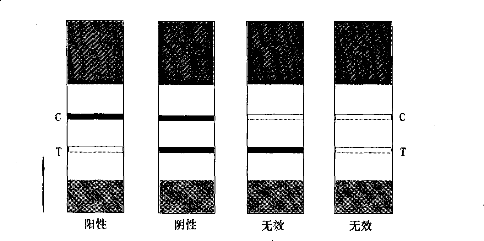
[0060] Prepare kanamycin, gentamicin, neomycin, streptomycin, chloramphenicol, cephalexin, sulfadimethoxine, and enrofloxacin into a sample with a concentration of 10ng/mL, and drop it on Test on the sample pad or sample hole. The test results (see Table 1) show that the kanamycin sample detection line has no color, and a red band appears on the quality control line, while gentamicin, neomycin, streptomycin, chloramphenicol, and cephalexin , Sulfadimethoxine, enrofloxacin sample test strip detection line and quality control line have red bands, which indicates that gentamicin, neomycin, streptomycin, chloramphenicol, cephalexin, sulfa Dimethoxypyrimidine and enrofloxacin have no cross-reaction with the test strip of the present invention, which shows that the test strip of the present invention has good specificity.
[0061] Table 1 Test results of specificity of test strips of the present invention
[0062]
[0063] Note: "+" means cross reaction; "-" means no cross reaction.
[0064] 2. Sensitivity test
[0065] The test strips of the present invention were used to detect kanamycin standard substances with concentrations of 0, 1, 5, 10, 50, and 100 ng/mL respectively, and the results are shown in Figure 4. The color of the detection line of the 1ng/mL kanamycin standard is very different from that of the 0ng/mL kanamycin standard. When the concentration of the kanamycin standard is higher than 10ng/mL, the color of the detection line does not appear . Therefore, the detection test strip of the present invention has a sensitivity of 1 ng/mL, which meets the "residue limit standard of kanamycin" stipulated by the European Union.
[0066] 3. Repeatability test
[0067] Prepare three sample solutions with different concentrations of 1, 10, and 100ng/mL with kanamycin standards. Negative samples and samples to be tested are tested with the test strip of the present invention. Each concentration has 6 replicates, and the replicates are tested. The results are shown in Table 2. The detection results are completely consistent and the color rendering is uniform, which proves that the detection result of the detection test strip of the present invention is stable and reliable, and has good repeatability.
[0068] Table 2 Repeatability test results of test strips of the present invention
[0069]
[0070] Note: "+" means positive result; "-" means negative result.

PUM

no PUM

Description & Claims & Application Information

We can also present the details of the Description, Claims and Application information to help users get a comprehensive understanding of the technical details of the patent, such as background art, summary of invention, brief description of drawings, description of embodiments, and other original content. On the other hand, users can also determine the specific scope of protection of the technology through the list of claims; as well as understand the changes in the life cycle of the technology with the presentation of the patent timeline. Login to view more.

Similar technology patents

Classification and recommendation of technical efficacy words

  • High sensitivity
  • Strong specificity

System and method for optical coherence imaging

ActiveUS20060055936A1Sufficient signal to noise ratioHigh sensitivityInterferometersScattering properties measurementsPhysicsCoherent imaging
Owner:THE GENERAL HOSPITAL CORP

Catheter-free implantable needle biosensor

ActiveUS20080033269A1High sensitivitySimple stepSurgeryVaccination/ovulation diagnosticsNeedle electrodeElectricity
Owner:VIRTUE MEDICAL TECH (HUZHOU) CO LTD

Cas9-foki fusion proteins and uses thereof

ActiveUS20150071899A1Strong specificityReduce the possibilityFusion with DNA-binding domainPeptide/protein ingredientsResearch settingNuclease
Owner:PRESIDENT & FELLOWS OF HARVARD COLLEGE

c-MET MODULATORS AND METHOD OF USE

InactiveUS20070179130A1Strong specificityEliminate side effectsBiocideSenses disorderSignal transductionCellular activity
Owner:EXELIXIS INC
Why Eureka
Try Eureka