Heterogeneous dermis reticular layer stent without basement membrane and cell as well as preparation method thereof

A mesh layer and basement membrane technology, applied in the field of medical biomaterials, can solve the problems of incomplete cell elution, low survival rate, poor nutrient permeability, etc. Effect

Inactive Publication Date: 2009-08-05
THE FIRST AFFILIATED HOSPITAL OF THIRD MILITARY MEDICAL UNIVERSITY OF PLA
View PDF2 Cites 9 Cited by
  • Summary
  • Abstract
  • Description
  • Claims
  • Application Information

AI Technical Summary

Problems solved by technology

These methods are difficult to remove immunogenic substances, and there are still varying degrees of immune rejection after transplantation
In the prior art, there is a piece of CN1460527A disclosed on December 10, 2003, which uses glutaraldehyde as a chemical cross-linking agent to reduce the immunogenicity of the dermis, but thus changes the natural structure of the dermis fibers, making the dermis more dense, which affects the implantation survival rate; another CN1343523A disclosed on April 10, 2002, which utilizes laser drilling to increase the porosity and permeability of the scaffold, but the pores obtained in this way are not the natural structure of fibers, Moreover, the laser has a thermal denaturation effect on the collagen fibers around the hole.
The various heterogeneous decellularized dermis of the prior art still have the following disadvantages: (1) the cell elution is not complete, the immunogenicity is strong, and local inflammatory immune reactions exist for a long time; poor permeability and slow vascularization, resulting in poor survival after transplantation

Method used


Image

  • Heterogeneous dermis reticular layer stent without basement membrane and cell as well as preparation method thereof
  • Heterogeneous dermis reticular layer stent without basement membrane and cell as well as preparation method thereof
  • Heterogeneous dermis reticular layer stent without basement membrane and cell as well as preparation method thereof

Examples


Embodiment 1

[0042] Using rats as the material source, the specific preparation process steps are as follows:

[0043] 1. Aseptically cut the back skin and subcutaneous tissue of rats;

[0044] 2. Soak in 0.1% bromogeramine solution for 20 minutes;

[0045] 3. Wash three times with phosphate buffer (pH7.2);

[0046] 4. Use a sterile skin extractor to remove the subcutaneous tissue to obtain full-thickness skin with a thickness of 0.5mm;

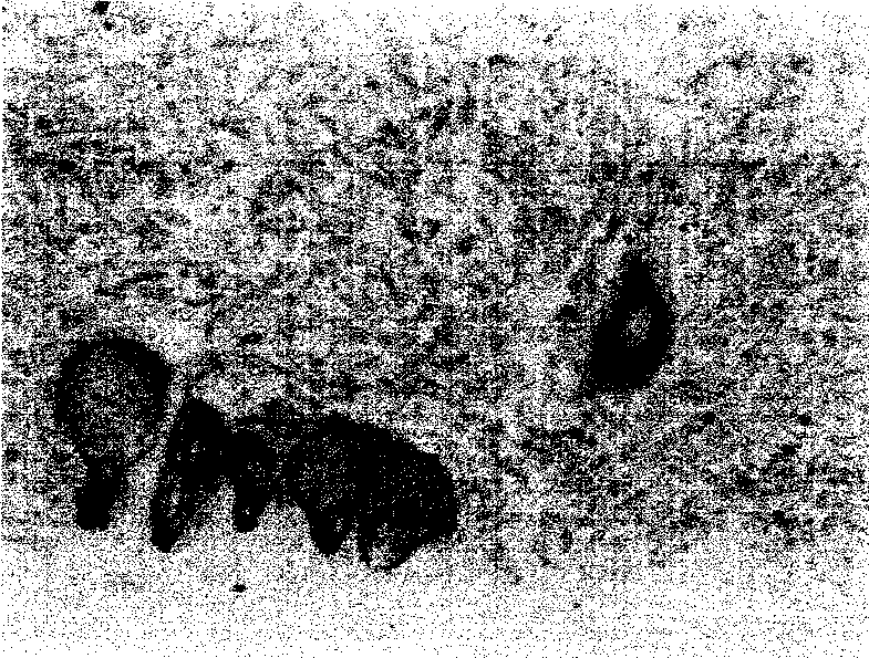
[0047] 5. Remove the epidermis, basement membrane and papillary dermis together with a sterile skin extractor to obtain a dermal reticular layer with a thickness of 0.3 mm (see figure 1 );

[0048] 6. Soak the obtained dermal reticular layer with hypotonic distilled water and continue to shake at 130 times / min for 1 hour;

[0049] 7. Use the digestion solution with a weight concentration of 0.25% trypsin and 0.02% EDTA·Na2, and shake continuously at 37°C for 3 hours;

[0050] 8. Replace the digestive solution with phosphate buffered saline, continue s...

Embodiment 2

[0055] Rabbits are used as the material source, and the specific preparation process steps are as follows:

[0056] 1. Aseptically cut the back skin and subcutaneous tissue of the rabbit;

[0057] 2. Soak in 0.1% bromogeramine solution for 20 minutes;

[0058] 3. Wash three times with phosphate buffer (pH7.2);

[0059] 4. Use a sterile skin extractor to remove the subcutaneous tissue to obtain full-thickness skin with a thickness of 0.4cm;

[0060] 5. Remove the epidermis, basement membrane and papillary dermis with a sterile skin remover to obtain a reticular layer of dermis with a thickness of 0.2mm;

[0061] 6. Soak the obtained dermal reticular layer with hypotonic distilled water and continue to shake at 130 times / min for 1.5 hours;

[0062] 7. Use the digestion solution with a weight concentration of 0.2% trypsin and 0.015% EDTA·Na2 at 37°C for 3 hours;

[0063] 8. Replace the digestive solution with phosphate buffered saline, continue shaking for 10 minutes, and was...


PUM

PropertyMeasurementUnit
thicknessaaaaaaaaaa

Abstract

The invention discloses an xenogenic dermal reticular layer scaffold with basilar membrane removal and no cells, which is taken from an dermal reticular layer of a mammal, and has a natural three-dimension structure with larger pores of the dermal reticular layer; the dermal scaffold removes a basilar membrane and has no cells; and the thickness of the dermal scaffold is between 0.2 and 0.8mm. The scaffold is prepared by performing sterile dermis getting, then removing subcutaneous tissue, epidermis, the basilar membrane and a dermal papillary layer, then orderly using distilled water to soak and using trypsinase and an EDTA.Na2 digestive juice to digest, using a phosphate buffer solution to wash after the persistent oscillation, then placing into a Triton X-100 solution for soaking and performing the persistent oscillation, and then using the phosphate buffer solution to wash. The scaffold completely removes the source of a main immunogenic substance in dermis, and reduces the immunogenicity; the scaffold has strong nutrition permeability, quick vascularization speed, high survival rate and good effect when used for the repair of skin wounds, the construction of tissue engineering skin, and tissue filling; and the scaffold has the advantages of rich dermis sources, simple preparation technology, short production period, and low cost.

Description

Technical field: [0001] The invention belongs to medical biomaterials, in particular to a heterogeneous dermis reticular layer scaffold used for skin wound repair, tissue engineering skin construction and tissue filling, and a preparation method thereof. Background technique: [0002] Clinically, a variety of injuries and diseases can lead to skin defects, especially for patients with extensive burns. Early scab removal and timely closure of wounds with dressings are currently the most effective and critical measures for skin repair. The use of autologous skin grafts from patients may cause scar hyperplasia and pigmentation in the donor area; scar hyperplasia occurs to varying degrees after wound healing, and there is a serious shortage of autologous skin sources in patients with severe burns. Therefore, finding an ideal skin wound repair material has become the main topic of wound repair. In 1913, Loewe confirmed that dermal tissue can enhance the toughness of grafted skin...

Claims


Application Information

Patent Timeline
Patent Type & Authority Applications(China)
IPC IPC(8): A61L27/36A61L27/56A61L27/60
Inventor 彭代智郑必祥朱崇涛何斌杨燕妮
Owner THE FIRST AFFILIATED HOSPITAL OF THIRD MILITARY MEDICAL UNIVERSITY OF PLA
Why Eureka
Try Eureka