Hepatoxic substance sieving and evaluating method based on fluorescence labeling

An evaluation method, fluorescent labeling technology, applied in biochemical equipment and methods, fluorescence/phosphorescence, microbial measurement/inspection, etc., can solve the problem of high price and achieve reliable results

Inactive Publication Date: 2010-07-28
ZHEJIANG UNIV
View PDF0 Cites 21 Cited by
  • Summary
  • Abstract
  • Description
  • Claims
  • Application Information

AI Technical Summary

Problems solved by technology

However, commercial HCS equipment is often very expensive, and only large pharmaceutical companies have the a

Method used


Image

  • Hepatoxic substance sieving and evaluating method based on fluorescence labeling
  • Hepatoxic substance sieving and evaluating method based on fluorescence labeling
  • Hepatoxic substance sieving and evaluating method based on fluorescence labeling

Examples


Example Embodiment

[0052] Example 1 Construction and application of screening system for liver toxicity screening

[0053] 1. Hardware and software system

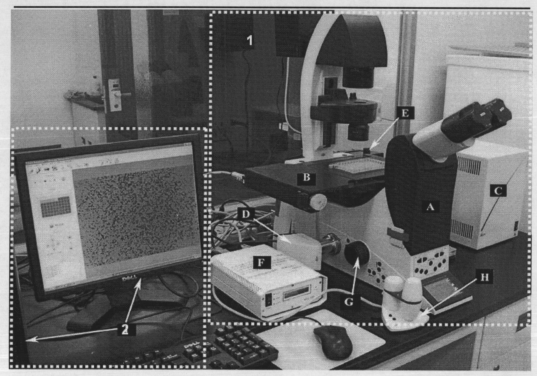
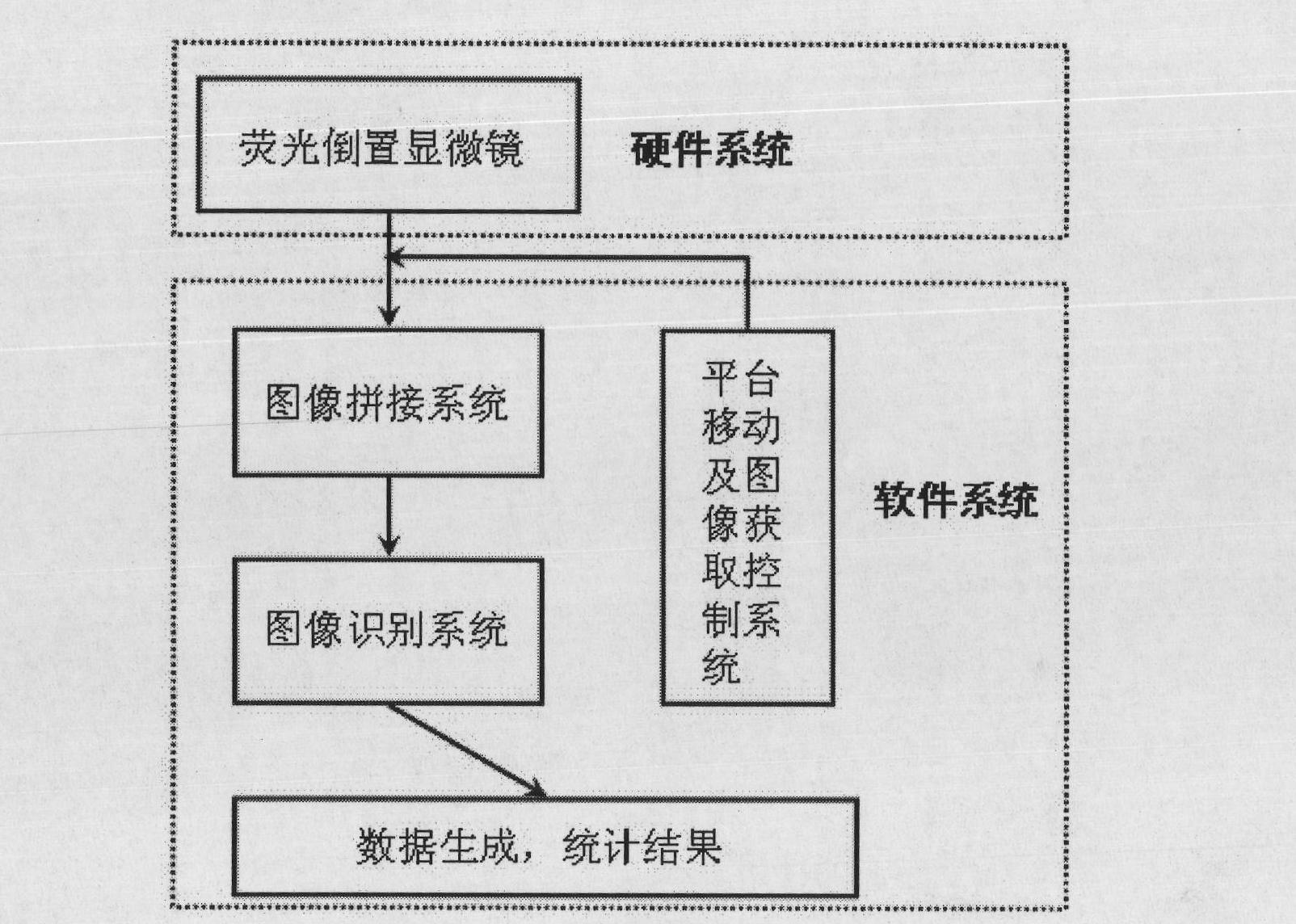
[0054] The purpose of the present invention is to provide a method for screening and evaluating hepatotoxic substances based on fluorescently labeled cell status, which is a combination of a hardware / software system for screening and evaluation. The screening and evaluation hardware system is powered by a German Leica DMI 6000B Fluorescence inverted microscope 1 and computer 2 constitute (see figure 1 ). Among them, the fluorescent inverted microscope 1 includes the fluorescent inverted microscope body A, high-precision controllable electric platform B, platform controller C, charge coupled device D (1392×1040 pixels, model Leica DFC 310FX, Leica company, Germany), mercury lamp E, mercury Lamp controller F, coarse adjustment screw G and manual joystick H, platform controller C is connected to microscope body A through wiring, and charge coupler ...

Example Embodiment

[0071] Example 2 Fluorescence image recognition processing

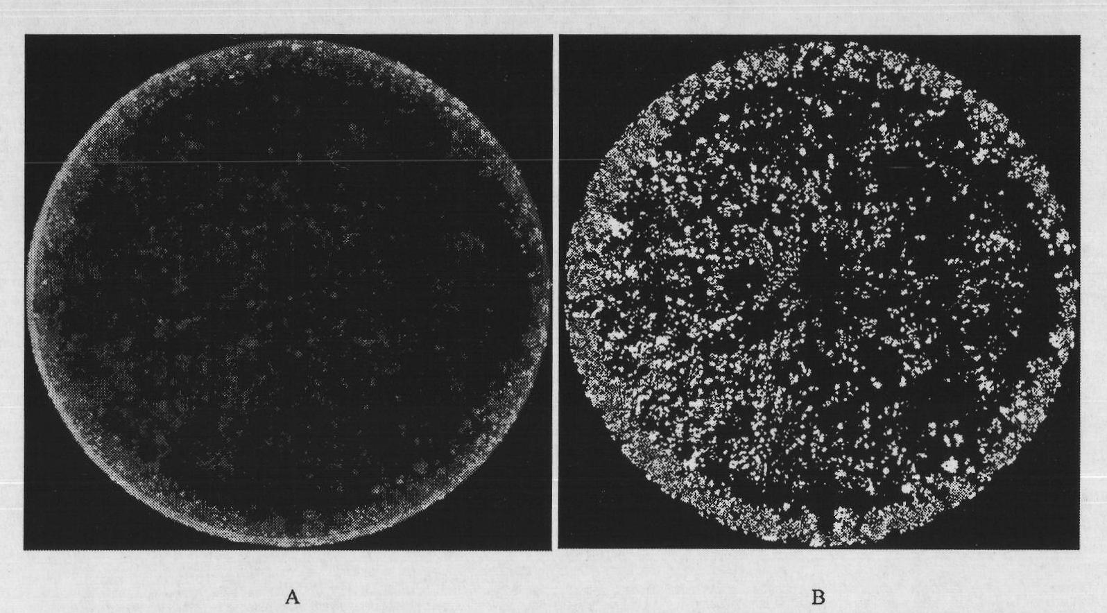
[0072] The fluorescence image recognition system is mainly to process the fluorescence images obtained by the fluorescence microscope, so that the biological information of the cells is converted into data, and the results are visualized. The specific processing steps are the deduction of the background signal of the fluorescent image, the binarization processing, and the data statistics generating report. Through the processing of these three steps, it is possible to visualize the fluorescent signal that cannot be distinguished by the human eye, with high recognition sensitivity, low detection limit, automatic batch processing of fluorescent pictures, and the result is faster, more stable, and multi-parameter ( Provide quantity, area and fluorescence intensity). See the identification diagram image 3 , Where Figure A. FDA stained whole-well HepG2 cells in a 96-well plate; Figure B. After image processing.

Example Embodiment

[0073] Example 3 FDA dye-labeled live cells based on the hardware and software system are used in the dose-effect test of the toxicity of two compounds, acetaminophen and chlorpromazine hydrochloride, on liver cells

[0074] HepG2 cells and L-O2 cells were seeded in a 96-well plate at a density of 3000 cells per well, and the medium was changed the next day. The compounds paracetamol and chlorpromazine hydrochloride were selected as positive injury controls, and a concentration gradient experiment was performed. Acetaminophen was diluted in half from 13.65 mM in 9 concentrations, and chlorpromazine hydrochloride was diluted in half in 9 concentrations from 163.5 μM, each with 3 replicate wells in parallel. The compound and the cells were incubated in a cell incubator for 48 hours. In a dark room, the culture medium in each well was discarded, and 100 μL / well of PBS containing 2.5 μg / mL FDA was added, and incubated at room temperature (25°C) for 15 minutes, followed by 100 μL / well....


PUM


Abstract

The invention provides a hepatoxic substance sieving and evaluating method based on cell states labeled by fluorescence, which is a sieving and evaluating method combining a hardware/software system. The images are acquired by a fluorescence inverted microscope and a computer in real time, the biological information is processed and visualized, the liner dynamic range width of the living cell is measured by combining FDA dyeing, and then the relevant parameters for evaluating the hepatoxic substances are provided. The method is quick, sensitive and economical, and is used for quickly detecting the influence of the sieved sample on cell quantity, forms, migration, apoptasis and the like under the condition of maintaining cell structure and function integrity, accurately carrying out quantitative analysis on the activity of the cell labeled by the fluorescence specialty and detecting the activity change of the cell under the function of the hepatoxic substances. The method has reasonable design. The provided sieving and evaluating system has complete structure, and can be used for sieving and evaluating the hepatoxic substances.

Description

technical field [0001] The invention belongs to the field of toxicological screening and evaluation methods of substances, and relates to the application of fluorescent probes to specifically mark cells, automatic acquisition technology of cell microscopic images, fluorescent image recognition and data generation, and to obtain hepatotoxic substance-related indicators by analyzing image information and use them in Hepatotoxicity screening and evaluation of exogenous substances, and this method can be extended to the screening and evaluation of other cytotoxic substances (such as nephrotoxicity, cardiotoxicity, anti-tumor, etc.). Background technique [0002] The liver is the most important place for the metabolism of drugs and exogenous substances in the body. While many drugs play a role in disease prevention and treatment in the body, they will inevitably affect the structure and function of the liver, leading to drug-induced liver injury of different mechanisms. According...

Claims


Application Information

Patent Timeline
IPC IPC(8): G01N21/64C12Q1/02
Inventor 程翼宇范骁辉王毅瞿海斌
Owner ZHEJIANG UNIV
Why Eureka
Try Eureka