

Duck plague virus cyst membrane gI protein polyclonal antibody as well as preparation method and application thereof
A polyclonal antibody, duck plague virus technology, applied in the fields of biotechnology and animal immunology, can solve problems such as lag, and achieve the effects of high specificity, high titer and simple operation
- Summary
- Abstract
- Description
- Claims
- Application Information
AI Technical Summary
Problems solved by technology
Method used
Image

Examples
Embodiment 1
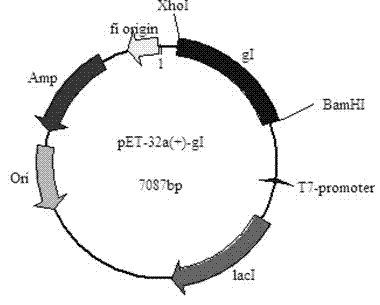
[0033] 1. Preparation of duck plague virus envelope gI protein
[0034] 1. Proliferation and Culture of Duck Plague Virus
[0035] Inoculate the CHv strain of duck plague virus into the newly grown dense monolayer duck embryo fibroblasts (DEF), discard the virus solution after adsorption at 37°C for 60 minutes, and then add calf serum with a volume fraction of 2% and 100IU / mL double Anti-constituent MEM was maintained in nutrient solution, followed by incubation at 37°C for 48 hours.
[0036] 2. DNA extraction
[0037] 1) Select DEF (100mL cell bottle) with 60%-70% cytopathic effect (CPE) after infection with DPV seed virus;
[0038] 2) Pour off the cell culture medium, add 500 μL of cell lysate, and at the same time add proteinase K (10 mg / mL) to a final concentration of 200 μg / mL, mix gently, and incubate at 37°C for 10 minutes;
[0039] 3) Pour the cell suspension into an EP centrifuge tube, wash the lysate remaining in the cell bottle with 500 μL of saturated phenol, an...
Embodiment 2
[0082] Example 2 Preparation of Polyclonal Antibody Based on Recombinant Envelope gI Protein of Duck Plague Virus
[0083] 1. Preparation of recombinant protein vaccine
[0084] Mix paraffin oil and lanolin in a ratio of 3:1, heat to dissolve, and sterilize at high temperature to make incomplete Freund's adjuvant, and add 1 mg of BCG to each milliliter of incomplete adjuvant to make complete Freund's adjuvant. Before use, mix Freund's complete adjuvant or incomplete adjuvant with equal volumes of the antigen liquid, ultrasonically emulsify in an ice bath, and check the emulsification effect. The oil droplets are emulsified completely.
[0085] 2. The immunization procedure is as follows
[0086]
[0087] The day before the immunization, blood was drawn from the rabbits, and the serum was separated as a negative control. After the fourth immunization, the carotid artery was bled, and the blood was collected. Place at 37°C for 30 minutes, and freeze at 4°C overnight. T...
Embodiment 3
[0106] Example 3 Application of polyclonal antibody based on duck plague virus envelope gI protein in the preparation of duck plague virus and duck plague virus envelope gI protein indirect immunofluorescence detection reagent
[0107] The application of the polyclonal antibody of DPV gI protein in the indirect immunofluorescence method, the steps are as follows:
[0108] 1) Prepare duck embryo fibroblast fly sheets by conventional methods, inoculate DPV after DEF grows into typical spindle cells 24 hours later, and harvest cell fly sheets 24 hours after inoculation;
[0109] 2) Wash the residual nutrient solution on the surface with sterilized pre-cooled PBS;
[0110] 3) Fix with 4% paraformaldehyde at room temperature, wash with sterilized PBS for 3×10 minutes;
[0111] 4) Permeabilize with PBS with a mass fraction of 0.2% Triton for 15 minutes, wash with 0.1% Tween-20 PBS (PBST) (same as above);
[0112] 5) Block with PBST containing 5% bovine serum albumin (BSA) at 37°...
PUM
| Property | Measurement | Unit |
|---|---|---|
| Molecular weight | aaaaa | aaaaa |
Abstract
Description
Claims
Application Information