Method for preparing protein-loaded tissue engineering fiber support

A fiber scaffold and tissue engineering technology, applied in fiber processing, non-woven fabrics, textiles and papermaking, etc., can solve the problems of not mentioning protein conformational protection results, not achieving sustained release effect, etc., to improve the release curve and reduce aggregation. phenomenon, the effect of good biocompatibility

Inactive Publication Date: 2011-09-21
SHANGHAI JIAO TONG UNIV
View PDF2 Cites 18 Cited by
  • Summary
  • Abstract
  • Description
  • Claims
  • Application Information

AI Technical Summary

Problems solved by technology

The obtained in vitro release curve of the fiber has a relatively serious burst release phenomenon, with 27% released on the first

Method used


Image

  • Method for preparing protein-loaded tissue engineering fiber support
  • Method for preparing protein-loaded tissue engineering fiber support
  • Method for preparing protein-loaded tissue engineering fiber support

Examples


Embodiment 1

[0033] Fabrication and in vitro release of protein-loaded tissue engineering fibrous scaffolds by ordinary electrospinning

[0034] a) Dissolving BSA (0.5%, w / w) in 0.5 ml of an aqueous polymer polysaccharide (0.5%, w / w) solution as the inner water phase;

[0035] b) Poly(lactic-co-glycolic acid) copolymer (99%, w / w) was dissolved in 2.5 ml of an equal volume mixed organic solvent of N,N-dimethylformamide and chloroform as the external oil phase;

[0036] c) The inner water phase is added dropwise to the outer oil phase, and magnetically stirred at 200 rpm for 30 minutes to form a W / O emulsion.

[0037] d) Add the above-mentioned W / O emulsion into a syringe, apply high-voltage static electricity, use a micro-injection pump and a receiver, and electrostatically spin a fiber film at room temperature;

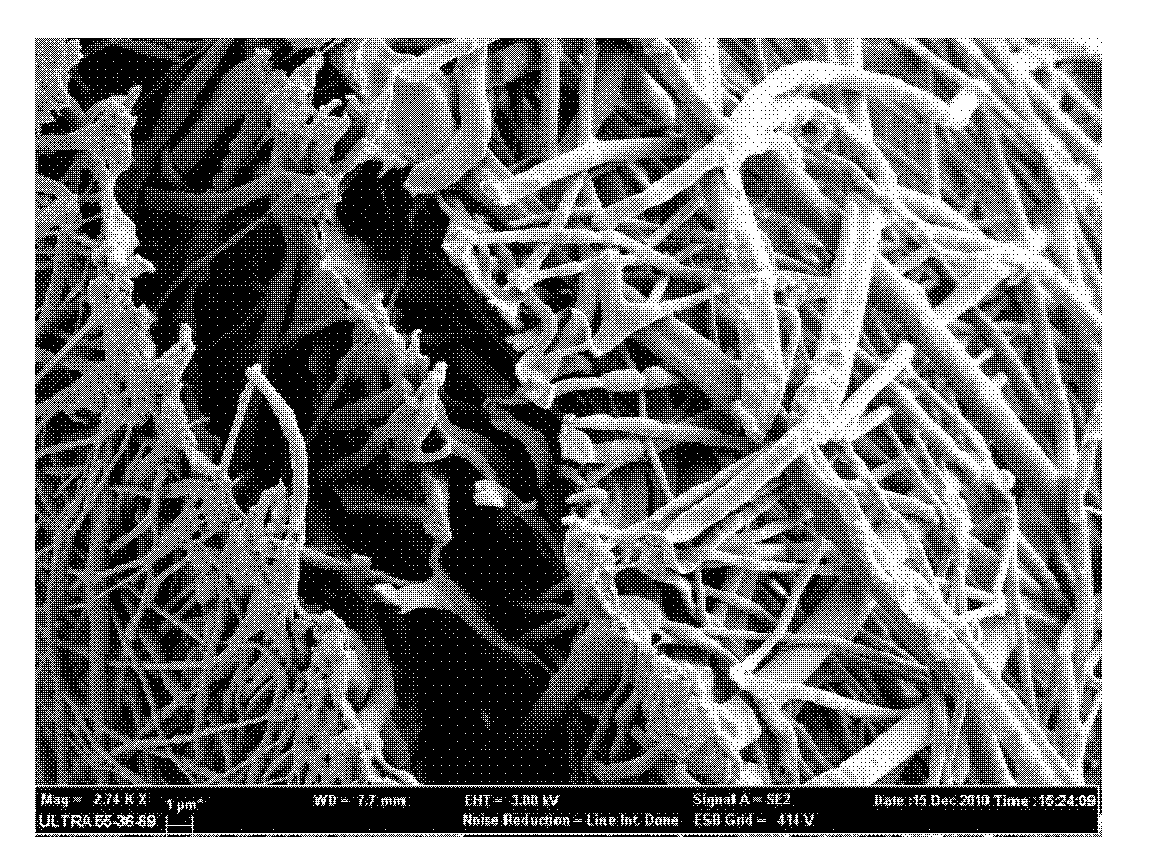
[0038] e) The fiber film is left to dry at room temperature to obtain tissue engineering fibers by emulsion electrospinning. The fiber has strong plasticity and can be molded in...

Embodiment 2

[0042] Fabrication of protein-loaded tissue engineering fibrous scaffolds by ordinary electrospinning and its release in vitro

[0043] a) Dissolving BSA (0.5%, w / w) in 0.5 ml of an aqueous polymer polysaccharide (0.5%, w / w) solution as the inner water phase;

[0044] b) Poly(lactic-co-glycolic acid) copolymer (99%, w / w) was dissolved in 10 ml of N,N-dimethylformamide and chloroform equal volume mixed organic solvent as the external oil phase;

[0045] c) The inner water phase is added dropwise to the outer oil phase, and magnetically stirred at 2500 rpm for 15 minutes to form a W / O emulsion.

[0046]d) Add the above-mentioned W / O emulsion into a syringe, apply high-voltage static electricity, use a micro-injection pump and a receiver, and electrostatically spin a fiber film at room temperature;

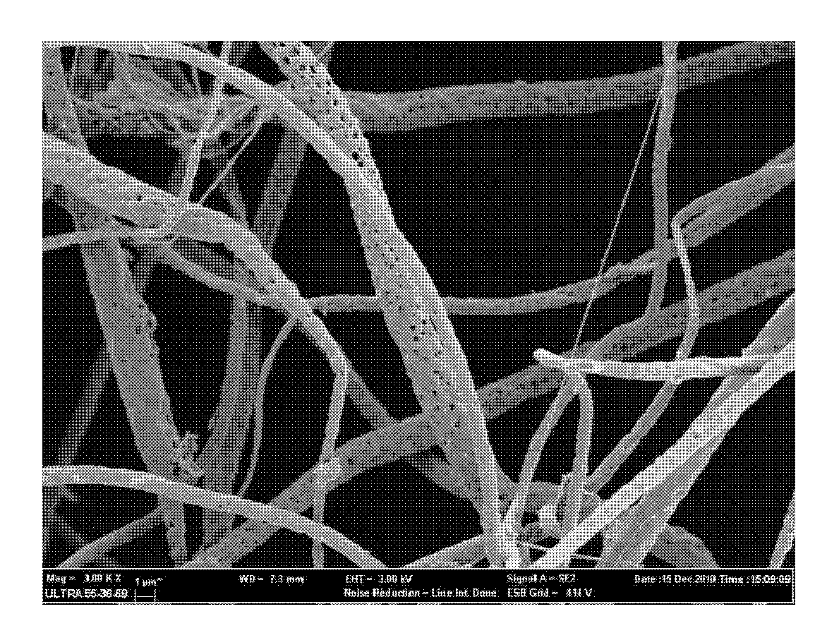
[0047] e) The fiber film is left to dry at room temperature to obtain tissue engineering fibers by emulsion electrospinning. The fiber has strong plasticity and can be molded into ...

Embodiment 3

[0049] Fabrication of protein-loaded tissue engineering fibrous scaffolds by ordinary electrospinning and its release in vitro

[0050] a) Dissolving BSA (5%, w / w) in 0.5 ml of an aqueous polymer polysaccharide (0.5%, w / w) solution as the inner water phase;

[0051] b) Poly(lactic-co-glycolic acid) copolymer (99%, w / w) was dissolved in 2.5 ml of an equal volume mixed organic solvent of N,N-dimethylformamide and chloroform as the external oil phase;

[0052] c) The inner water phase is added dropwise to the outer oil phase, and magnetically stirred at 2500 rpm for 15 minutes to form a W / O emulsion.

[0053] d) Add the above-mentioned W / O emulsion into a syringe, apply high-voltage static electricity, use a micro-injection pump and a receiver, and electrostatically spin a fiber film at room temperature;

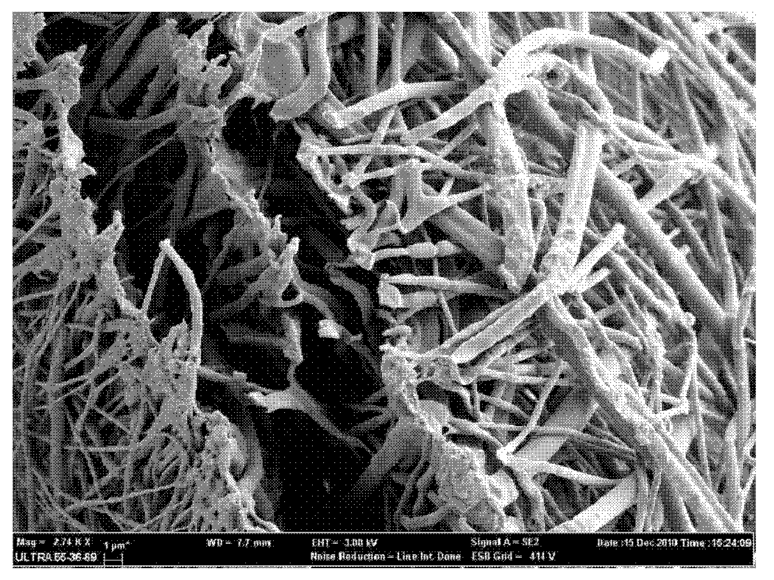
[0054] e) The fiber film is left to dry at room temperature to obtain tissue engineering fibers by emulsion electrospinning. The fiber has strong plasticity and can be molded...


PUM


Abstract

The invention relates to a method for preparing a protein-loaded tissue engineering fiber support in the technical field of medicaments, which comprises the following steps of: dissolving protein in macromolecular polysaccharide aqueous solution and using as an inner water phase; dissolving sustained-release macromolecules in an organic solvent, adding a suspending aid for dispersing, and using as an outer oil phase; adding the inner oil phase into the outer water phase dropwise, magnetically stirring or homogenizing to form emulsion, adding the emulsion into an injector, and performing electrostatic spinning by using a micro-injection pump and a receiver at room temperature under the condition of applying high-voltage static electricity to form a fiber membrane, or respectively putting the inner water phase and the outer oil phase into a capillary tube subjected to coaxial cospinning, and performing the electrostatic spinning by using the micro-injection pump and the receiver at room temperature under the condition of applying the high-voltage static electricity on inner and outer layers simultaneously; and standing and airing at room temperature to obtain tissue engineering fibers. The tissue engineering fiber support comprises the following components in percentage by weight: 0.5 to 20 percent of protein or polypeptide medicaments, 2 to 20 percent of macromolecular polysaccharides, 0 to 15 percent of suspending aid and 60 to 99 percent of sustained-release macromolecules. By the method, release curves of the protein are improved, the stability in the processes of preparation, storage and release is improved, and the medicament loading capacity of the fibers is increased.

Description

technical field [0001] The present invention relates to a method for preparing a tissue engineering fiber scaffold in the field of pharmaceutical technology, in particular to a method for preparing a protein-loaded tissue engineering fiber scaffold. Background technique [0002] At present, in the preparation method of emulsion-based electrospinning tissue engineering fiber scaffolds, the problem of protecting the activity and conformation of protein drugs is usually ignored, and there is also the phenomenon of initial burst release of protein or polypeptide drugs. [0003] Found through literature search to prior art, Li Xiaoqiang etc. are in Colloids and Surfaces B: Biointerfaces 2010 the 75th phase 418-424 pages, article title "Encapsulation of proteins in poly(l-lactide-co-caprolactone) fibers byemulsion electrospinning "(Polylactic acid-caprolactone copolymer fiber loaded with protein prepared by emulsion electrospinning), proposed to use bovine serum albumin (BSA, 7.69...

Claims


Application Information

Patent Timeline
IPC IPC(8): A61L27/54A61L27/20A61L27/18A61L31/16A61L31/06A61L31/04D04H1/72D01D5/00D01D5/30D01F6/92D01F1/10
Inventor 吴飞金拓袁伟恩高小寒魏蓓蓓秦明杰
Owner SHANGHAI JIAO TONG UNIV
Why Eureka
Try Eureka