Application of H22 liver cancer cell autophagosome to preparation of liver cancer therapeutic vaccine

A therapeutic vaccine and liver cancer cell technology, applied in the field of liver cancer vaccine development to achieve the effect of inhibiting the growth of tumors

Active Publication Date: 2012-05-02
SOUTHEAST UNIV
View PDF0 Cites 8 Cited by
  • Summary
  • Abstract
  • Description
  • Claims
  • Application Information

AI Technical Summary

Problems solved by technology

However, there are many problems to be solved in research experiments, including the source of DC, the preparation process, the antigen load of DC, the dosage, the frequency of use, the route and frequency of immunization, and how to better recruit tumor antigens.

Method used


Image

  • Application of H22 liver cancer cell autophagosome to preparation of liver cancer therapeutic vaccine
  • Application of H22 liver cancer cell autophagosome to preparation of liver cancer therapeutic vaccine
  • Application of H22 liver cancer cell autophagosome to preparation of liver cancer therapeutic vaccine

Examples


Embodiment 1

[0039] Example 1: Preparation of autophagosome DRibbles in H22 / BNL liver cancer cells.

[0040] 1. Culture of H22 / BNL liver cancer cells:

[0041]Take the H22 / BNL cells frozen in liquid nitrogen, add 1640 culture medium containing 10% (V / V) fetal bovine serum, 100U / ml penicillin and 100U / ml streptomycin immediately after thawing in a water bath at 40°C, and resuspend the cells to 1×10 6 individual / ml, and transfer it into a 500ml cell culture flask, at 37°C, 5% (V / V) CO 2 Cultured in a constant temperature incubator, the medium was changed every 1 to 2 days, and routinely digested and passaged with 0.25% trypsin;

[0042] 2. Treatment of H22 / BNL liver cancer cells:

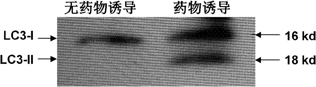
[0043] After the cells cultured in step (1) adhere well to the wall and have a moderate density, add 100 nmol / L of rapamycin, 200 nmol / L of Velcade, and ammonium chloride (NH 4 CL) 30mmol / L combined treatment for 16 hours, induced autophagosomes - DRibbles. And set up a control group (only adding culture medi...

Embodiment 2

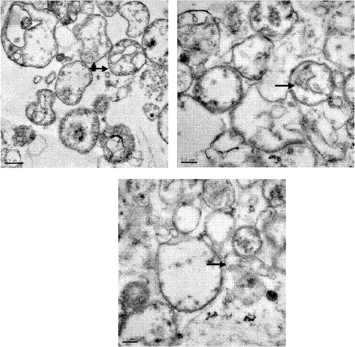
[0052] Example 2: Transmission Electron Microscopy Identification of Morphology of DRibbles.

[0053] The extracted DRibbles were centrifuged at high speed to compress them tightly, the supernatant was discarded, the precipitate was fixed with 2.5% (V / V) glutaraldehyde, and sent to the electron microscope room for processing, and the morphology of DRibbles was observed on the microscope. Such as figure 1 Shown: Under the electron microscope, small bodies with a membrane structure can be seen, with an average diameter of about 200-300 nm (pointed by the arrow), which proves that the autophagosomes of H22 liver cancer cells are effectively recruited.

Embodiment 3

[0054] Example 3: Determination of total protein content in DRibbles (Bradford method).

[0055] 1) Completely dissolve the protein standard, take 25 μl and dilute to 100 μl with PBS;

[0056] 2) Add 0, 1, 2, 4, 8, 12, 16, and 20 μl of the standard into the 96-well plate, and add PBS to make up to 20 μl;

[0057] 3) Freeze and thaw the DRibble to be tested repeatedly in advance, centrifuge to take the supernatant, add an appropriate volume of sample to a 96-well plate, and add PBS to make up to 20 μl;

[0058] 4) Add 200 μL of LG250 staining solution to each well, and place at room temperature for 3-5 minutes;

[0059] 5) Measure A595 with a microplate reader, or the absorbance of other wavelengths between 560-610nm;

[0060] 6) Use the software ElisaCalc to draw a standard curve, and calculate the protein concentration in the sample according to the standard curve, and the result is about 1-3 μg / μl.


PUM

PropertyMeasurementUnit
diameteraaaaaaaaaa
diameteraaaaaaaaaa

Abstract

The invention discloses application of H22 liver cancer cell autophagosome DRibbles to the preparation of a liver cancer therapeutic vaccine. Liver cancer is one of the most common malignant tumors in China and can be effectively prevented by developing a liver cancer vaccine; and years of research results prove that tumor cell autophagosome is one of effective vectors for tumor antigen cross-presentation. The application comprises a core technology for extracting the autophagosome DRibbles from human liver cancer cells and proves that the autophagosome can effectively induce anti-tumor immune response of organisms and has a good biotherapy prospect.

Description

technical field [0001] The invention belongs to the technical field of liver cancer vaccine development, and in particular relates to the application of H22 liver cancer cell autophagosomes in the preparation of liver cancer therapeutic vaccines. Background technique [0002] Hepatocellular carcinoma (HCC) is one of the malignant tumors with extremely high degree of malignancy and poor prognosis, which seriously threatens human health. Curative resection or liver transplantation is currently the preferred treatment for liver cancer patients, but due to factors such as multiple intrahepatic lesions, extrahepatic metastasis, and lack of donors, only 10% to 15% of newly diagnosed liver cancer patients can undergo surgery treat. Clinical data show that most liver cancer patients are not sensitive to chemotherapy, radiotherapy and other treatments. Therefore, the clinical treatment of liver cancer urgently needs new methods and means. As a new treatment strategy for liver cance...

Claims


Application Information

Patent Timeline
Patent Type & Authority Applications(China)
IPC IPC(8): A61K39/00C12N5/09A61P35/00
Inventor 王立新曹萌
Owner SOUTHEAST UNIV
Why Eureka
Try Eureka