Stent for osteochondral defect repair and preparation method thereof

An osteochondral and cartilage layer technology, applied in medical science, prosthesis, etc., can solve problems such as the need to further improve the mechanical properties of the scaffold, increase the manufacturing difficulty and cost, and block the natural transition between bone and cartilage, so as to achieve good osteochondral defects. Repair effect, easy mass production, low production technical requirements

Inactive Publication Date: 2012-07-04
WEST CHINA HOSPITAL SICHUAN UNIV
View PDF7 Cites 19 Cited by
  • Summary
  • Abstract
  • Description
  • Claims
  • Application Information

AI Technical Summary

Problems solved by technology

This technical solution has the following problems: 1. Since the scaffold is composed of cartilage layer, calcification layer and subchondral bone layer from top to bottom, the calcification layer is a prefabricated cartilage calcification layer, which will block the natural transition between bone and cartilage, because normal cartilage The calcified layer is a cell-matrix complex formed by the natural activities of living chondrocytes under the action of biomechanics and local microenvironment
2. The support has a three-layer structure, and its structure is complicated, which increases the difficulty and cost of production
3. The mechanical properties of the scaffold need to be further improved
4. The use of 3D printing technology requires high technical requirements, which is not conducive to large-scale promotion

Method used


Image

  • Stent for osteochondral defect repair and preparation method thereof
  • Stent for osteochondral defect repair and preparation method thereof
  • Stent for osteochondral defect repair and preparation method thereof

Examples


Embodiment 1

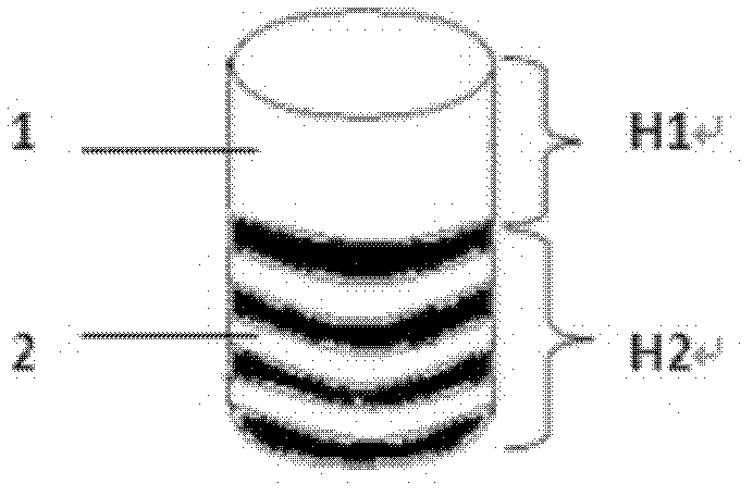
[0037] In this embodiment, the scaffold used for osteochondral defect repair is as figure 1 As shown, it is a cylinder with a diameter of 3 mm, consisting of a cartilage layer 1 and a subchondral bone layer 2, the thickness H1 of the cartilage layer 1 is 20 mm, the thickness H2 of the subchondral bone layer 2 is 25 mm, the cartilage layer 1 and the subchondral bone layer The subchondral bone layer 2 is closely integrated. The material of cartilage layer 1 is a mixture of type I collagen and chondroitin sulfate, the mass ratio of type I collagen to chondroitin sulfate is 20:1, and the material of subchondral bone layer 2 is porous titanium-chromium alloy with multiple composite pores. The diameter of the large pores is 7000-10000 μm, the diameter of the small pores is 700-1000 μm, and the porosity is 85%.

[0038] The processing steps of the preparation method are as follows:

[0039] (1) Preparation of subchondral bone layer

[0040] Machining a porous titanium-chromium all...

Embodiment 2

[0050] In this embodiment, the scaffold used for osteochondral defect repair is as figure 1 As shown, it is a cylinder with a diameter of 20 mm, consisting of a cartilage layer 1 and a subchondral bone layer 2, the thickness H1 of the cartilage layer 1 is 5 mm, the thickness H2 of the subchondral bone layer 2 is 3 mm, the cartilage layer 1 and the subchondral bone layer The subchondral bone layer 2 is closely integrated. The material of cartilage layer 1 is a mixture of type I collagen and chondroitin sulfate, the mass ratio of type I collagen to chondroitin sulfate is 5:1, and the material of subchondral bone layer 2 is porous titanium with multiple composite pores. The pore diameter is 1000-3000 μm, the small hole diameter is 10-300 μm, and the porosity is 55%. The process steps are as follows:

[0051] (1) Preparation of subchondral bone layer

[0052] Machining porous titanium with multiple composite pores to the shape and size of the subchondral bone layer 2 as describe...

Embodiment 3

[0062] In this embodiment, the scaffold used for osteochondral defect repair is as figure 1 As shown, it is a cylinder with a diameter of 6 mm, consisting of a cartilage layer 1 and a subchondral bone layer 2, the thickness H1 of the cartilage layer 1 is 3 mm, the thickness H2 of the subchondral bone layer 2 is 6 mm, the cartilage layer 1 and the subchondral bone layer The subchondral bone layer 2 is closely integrated. The material of cartilage layer 1 is a mixture of type I collagen and chondroitin sulfate, the mass ratio of type I collagen to chondroitin sulfate is 5.63:1, and the material of subchondral bone layer 2 is porous titanium oxide with multiple composite pores. The pore diameter is 1000-7000 μm, the small pore diameter is 100-700 μm, and the porosity is 70%. The process steps are as follows:

[0063] (1) Preparation of subchondral bone layer

[0064] processing porous titanium oxide with multiple composite pores into the shape and size of the subchondral bone l...


PUM

PropertyMeasurementUnit
thicknessaaaaaaaaaa
thicknessaaaaaaaaaa
pore sizeaaaaaaaaaa

Abstract

The invention discloses a stent for osteochondral defect repair. The stent consists of a cartilage layer with pores and a subchondral bone layer with pores, wherein the cartilage layer and the subchondral bone layer are tightly combined into a whole; the material of the cartilage layer is a mixture of type I collagen and chondroitin sulfate; and the material of the subchondral bone layer is porous titanium or porous titanium oxide or a porous titanium alloy. A preparation method of the stent comprises the following steps of: (1) processing the porous titanium or porous titanium oxide or porous titanium alloy to a shape and a size required by the subchondral bone layer; (2) preparing cartilage layer slurry; and (3) molding and treating a stent, putting the subchondral bone layer prepared in the step (1) into a mold, injecting the cartilage layer slurry prepared in the step (2) into the mold, putting the mold into a freeze dryer, freezing for at least 5 hours, molding, performing thermal crosslinking and chemical crosslinking on the molded stent, and putting the chemically-crosslinked stent into sterile water for rinsing to obtain the stent for osteochondral defect repair.

Description

technical field [0001] The invention belongs to components for repairing osteochondral defects, in particular to a bracket for repairing osteochondral defects. Background technique [0002] Cartilage damage is very common in orthopedic diseases, but articular cartilage damage is difficult to treat. In clinic and experiment, "scaffold" is used for the repair of osteochondral defect. [0003] CN 101219069A discloses a double-layer composite support for osteochondral repair, which is composed of a base frame, one end of which is a simulated cartilage-like layer, and the other end is a simulated subchondral bone layer, the two layers are tightly combined, located The outer surface of the simulated subchondral bone layer is provided with threads. The material for simulating the cartilage-like layer is one or a mixture of chitin, chitin collagen, fibrin, alginate, silk protein, polylactic acid, polyglycolic acid, polylactic acid-polyglycolic acid copolymer; The material for sim...

Claims


Application Information

Patent Timeline
Patent Type & Authority Applications(China)
IPC IPC(8): A61L27/40A61L27/26A61L27/06A61L27/02A61L27/56
Inventor 项舟段鑫吕晶同
Owner WEST CHINA HOSPITAL SICHUAN UNIV
Why Eureka
Try Eureka