Optical clearing agent for bone tissue

A light-transparent agent and bone tissue technology, applied in the field of biomedical optical imaging, can solve the problems of hard bone tissue transparency, restricting the development and application of cortical optical imaging for transcranial living imaging, and achieving improved imaging depth and reduced tissue internal scattering Effect

Active Publication Date: 2014-07-09
HUAZHONG UNIV OF SCI & TECH
View PDF1 Cites 1 Cited by
  • Summary
  • Abstract
  • Description
  • Claims
  • Application Information

AI Technical Summary

Problems solved by technology

However, due to the differences in the composition and structure of soft tissue and bone tissue, these light clearing agents that have been proven to be equally effective for soft tissue cannot make hard bone tissue transparent, which severely restricts the development and application of cortical optical imaging for transcranial live imaging.

Method used


Image

  • Optical clearing agent for bone tissue
  • Optical clearing agent for bone tissue
  • Optical clearing agent for bone tissue

Examples


Embodiment 1

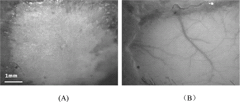
[0015] The scalp of a C57 mouse (20 weeks old) was cut open to expose a skull of about 1 cm × 1 cm square. According to the ratio of each component of the solution in Example 1 in Table 1, the optical transparent agent is configured into a mixed solution, which is directly added dropwise to the skull of the living mouse, and spread evenly (0.5-0.8ml / cm 2 ). figure 1 The CCD images taken before and after the treatment of the living mouse skull with light clearing agent are given. in figure 1 (A) The skull of a normal C57 mouse is exposed. Due to the opacity of the skull, it is impossible to see the intracranial cortical blood vessels; figure 1 (B) 5 minutes after adding the optical clearing agent, the skull becomes transparent to light, the cortical blood vessels below the skull become clearly visible, and some small branches can also be distinguished in the field of vision.

Embodiment 2

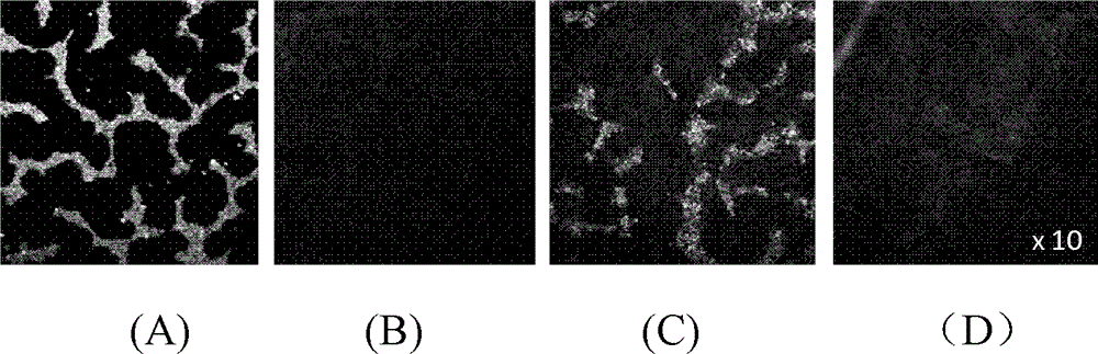
[0017] The isolated skull was taken from a 4-week-old SD rat, which was covered on the encapsulated fluorescent beads solution, and observed with a fluorescent microscope. The optical transparent agent is configured according to the ratio of the components of the solution in Example 2 in Table 1, and the rat skull is soaked in vitro (1.5-2ml / cm 2 ) in the light clearing agent, and after 5 minutes, cover the encapsulated fluorescent beads solution for imaging. The light-clearing effect can be reversed by flushing the skull treated with light-clearing agent with saline, and then imaging under a fluorescent microscope. figure 2 The fluorescence signals observed without skull, with skull, after light transparency of skull and after reversal of light transparency with saline are given respectively. figure 2 (A) shows the fluorescence signal observed without the skull, and the fluorescence information at this time is very strong; from figure 2 (B) It can be seen that the skull ...

Embodiment 3

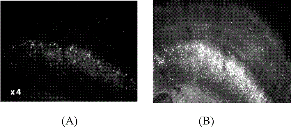
[0019] Cut the brain slices of GFP-labeled transgenic mice to a thickness of 100 μm. One group was not treated with optical clearing agent and observed directly under a fluorescent microscope; solution, directly applied to mouse brain slices (0.2-0.4ml / cm 2 ), observed with a fluorescence microscope after 1 min. image 3 Respectively given are before the action of the light clearing agent under the 4 times objective lens ( image 3 (A)) after ( image 3 (B) Fluorescence signal of mouse brain slices. Only a small amount of neuron cell bodies could be observed in rat brain slices that were not treated with light clearing agent; however, after short-term treatment with light clearing agent, the cell bodies of nerve cells became obviously brighter, and the dendrite structure was now clearly identifiable.

[0020] The different optical clarity agent formulations shown in Table 1 can be formulated and used according to the methods shown in Examples 1 to 3 above to achieve these e...


PUM


Abstract

The present invention discloses an optical clearing agent for bone tissue. The optical clearing agent comprises dimethyl sulfoxide, sodium bicarbonate, and at least two materials selected from polyethylene glycol-400, methyl salicylate, ethylenediaminetetraacetic acid, sodium diatrizoate, glycerol, glucose, sodium dodecylbenzene sulfonate, sorbitol and laurinol, wherein a volume percentage of the dimethyl sulfoxide in the optical clearing agent for bone tissue is 40-80%, a mass percentage of the sodium bicarbonate in the optical clearing agent for bone tissue is 1-2%, the balance is other materials, and the pH value of the optical clearing agent for bone tissue is 6-11. After the optical clearing agent for bone tissue in the present invention acts on bone tissue, a refractive index inside the tissue can be quickly homogenized, and scattering inside the tissue can be reduced, such that the tissue provides improved transparency for light, the imaging depth is increased, and a new method for obtaining cortex neuron subcellular structures and microvascular information is provided. After the optical clearing agent for bone tissue is locally coated on the bone tissues or is adopted to soak the bone tissues, the bone tissue can provide improved transparency for light.

Description

technical field [0001] The invention belongs to the technical field of biomedical optical imaging, in particular to a bone tissue optical transparency agent, which is a mixed solution capable of changing the optical transparency of bone tissue. Background technique [0002] With the development of biomedical photonics, modern optical imaging technology provides a new means for high-resolution acquisition of three-dimensional microstructure of biological tissues at the molecular level. However, the high scattering of visible and near-infrared light by biological tissues limits the penetration depth of light, so that these techniques can only image superficial tissues, and the image contrast decreases significantly with imaging depth. The optical transparency technology of biological tissue proposed in recent years can effectively enhance the penetration depth of light in biological tissue by introducing hyperosmotic and high refraction chemical reagents into biological tissue...

Claims


Application Information

Patent Timeline
Patent Type & Authority Patents(China)
IPC IPC(8): G01N1/28G01N1/30A61K49/00
Inventor 张洋
Owner HUAZHONG UNIV OF SCI & TECH
Why Eureka
Try Eureka