Method for in-situ detecting of drug molecules in animal tissues by dual-beam laser mass-spectrography

A technology for in-situ detection of drug molecules, applied in measuring devices, analyzing materials, and analyzing materials through electromagnetic means, can solve problems such as inability to detect, and achieve the effects of improved applicability, simple operation, and short pretreatment time

Inactive Publication Date: 2013-01-16
SOUTH CHINA NORMAL UNIVERSITY
View PDF2 Cites 6 Cited by
  • Summary
  • Abstract
  • Description
  • Claims
  • Application Information

AI Technical Summary

Problems solved by technology

However, at present, the detection of methylene blue is still relatively small, mainly Raman spectr

Method used


Image

  • Method for in-situ detecting of drug molecules in animal tissues by dual-beam laser mass-spectrography
  • Method for in-situ detecting of drug molecules in animal tissues by dual-beam laser mass-spectrography
  • Method for in-situ detecting of drug molecules in animal tissues by dual-beam laser mass-spectrography

Examples


Embodiment 1

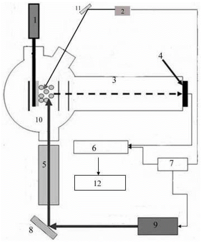
[0035] Embodiment 1: A kind of dual-beam laser mass spectrometer

[0036]The double-beam laser mass spectrometer comprises a digital delay generator 7, a sampling system 1, solid-state lasers 2 and 9, a vacuum system, a gas cell 5, a plano-convex mirror, a data acquisition device 6 and a data processing device 12; the vacuum system includes Ionization chamber 10, flight tube 3 and microchannel plate 4, one end of flight tube 3 is connected with ionization chamber 10, and the other end is provided with microchannel plate 4; Described solid-state laser is two, is placed next to the vacuum system respectively, solid-state laser 2 A B prism 11 is set between the ionization chamber 10, a gas cell 5 is set between the solid-state laser 9 and the ionization chamber 10, an A prism 8 is set between the solid-state laser 9 and the gas cell 5, and the side of the gas cell 5 close to the ionization chamber 10 A plano-convex mirror is set; the data acquisition device 6 is an oscilloscope, ...

Embodiment 2

[0037] Example 2: Using the dual-beam laser mass spectrometer described in Example 1, the detection of the photodynamic therapy drug molecule methylene blue in cancer tissue by dual-beam laser mass spectrometry.

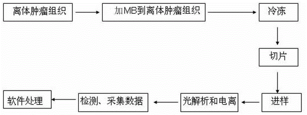
[0038] The steps of L2MS detection of methylene blue in cancer tissue are as follows ( figure 1 ):

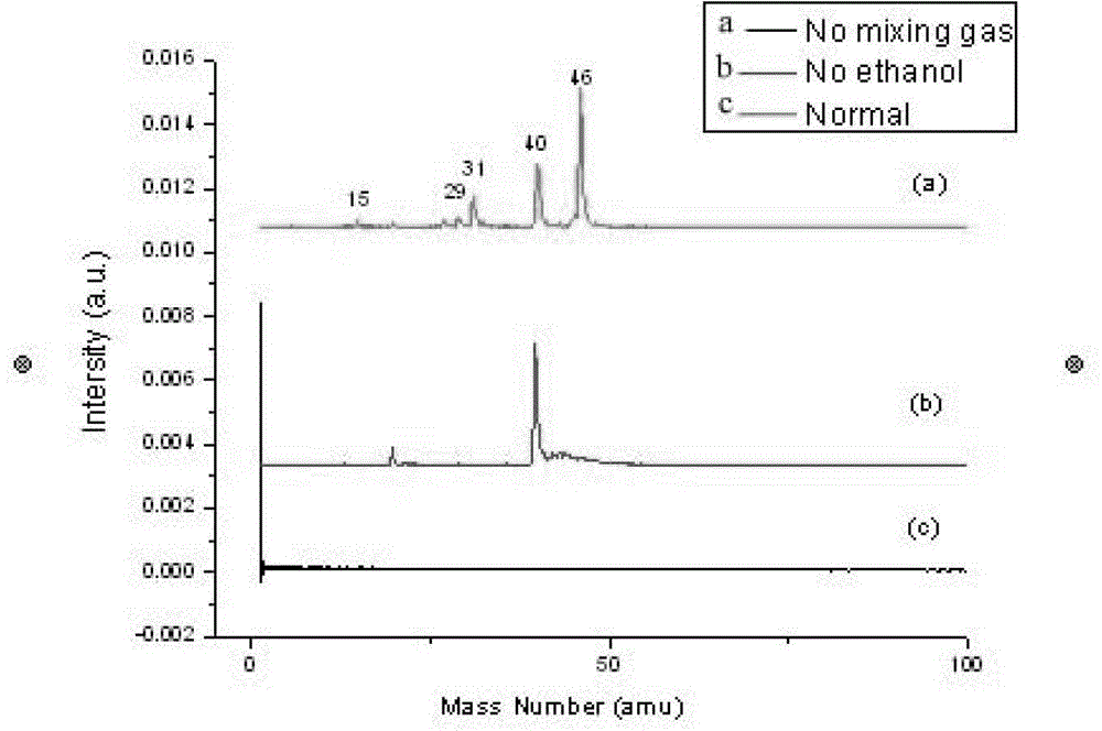
[0039] (1) Use ethanol as a test molecule to detect the performance of the entire mass spectrometry system

[0040] As a conventional laboratory reagent, ethanol has a low saturated vapor pressure. It can be cooled by combining ultrasonic molecular beam with helium as the carrier gas. The helium pressure is 0.1Mpa, and the vacuum degree of the molecular beam is 5.0×10 -3 Pa, and then ionize it with a vacuum ultraviolet laser, that is, a 118nm ionization laser, and the vacuum degree of the ionization chamber is 1.0×10 -5 Pa, where the delay time between the molecular beam and the ionizing light is 80μs, a relatively standard spectrum can be obtained to test the stabi...


PUM

PropertyMeasurementUnit
Thicknessaaaaaaaaaa
Wavelengthaaaaaaaaaa
Lengthaaaaaaaaaa

Abstract

The invention discloses a method for in-situ detecting of drug molecules in animal tissues by dual-beam laser mass-spectrography. The method comprises the steps that a drug molecular solution is added into isolated animal tumor tissues, and is frozen and sliced after being diffused adequately; the slices are fixedly arranged on graphite subjected to ultrasonic processing, the graphite is placed into a dual-beam laser mass-spectrometer, and the parsing laser emitted by the dual-beam laser mass-spectrometer enters into an ionization chamber after being focused by the parsing laser and is radiated on the slices; after delay of 18-30 microseconds, a second beam pulse laser is emitted, and is converted into a vacuum ultraviolet laser after being subjected to third harmonic generation in a gas pool; the second beam pulse laser is separated from the vacuum ultraviolet laser by a flat convex lens at a light outlet of the gas pool after the second beam pulse laser and the vacuum ultraviolet laser are focused; the focused vacuum ultraviolet laser enters into the ionization chamber in a direction parallel to a graphite rod and is intersected with the parsing laser; gasification-parsing and ionization are carried out on the isolated tumor tissues; ionized ions fly through a flight tube; and data collecting and processing are carried out.

Description

technical field [0001] The invention belongs to the detection technical field of small molecule drug development, and in particular relates to a method for detecting drug molecules in animal tissues using double-beam laser mass spectrometry (L2MS). Background technique [0002] Two-step Laser Desorption / Laser Ionization Mass Spectrometry (L2MS) uses two laser beams to complete the tasks of gasification / analysis and ionization respectively. L2MS has the following advantages: First, because the analysis laser and ionization laser and are separated in space, so the energy and wavelength of the two laser beams can be easily optimized independently according to actual needs; secondly, the experimental technique requires very little sample volume, and does not need to be artificially added to the sample to be tested Other chemical substances are used as the matrix, the sample preparation is simple, and the secondary pollution of the sample is avoided. Due to its unique advantages...

Claims


Application Information

Patent Timeline
IPC IPC(8): G01N27/64
Inventor 胡勇军王红磊杨晴官凯文李卫星
Owner SOUTH CHINA NORMAL UNIVERSITY
Why Eureka
Try Eureka