Real-time quantitative detection reagent and method of time-resolved fluorescence immune chromatography

A technology of test strips and kits, applied in the field of detection, can solve problems such as easy occurrence of false positives and affect the accuracy of test results, and achieve the effects of eliminating non-specific binding, improving signal-to-noise ratio and high specificity

Active Publication Date: 2013-01-16
上海执诚生物科技有限公司
View PDF7 Cites 45 Cited by
  • Summary
  • Abstract
  • Description
  • Claims
  • Application Information

AI Technical Summary

Problems solved by technology

[0004] Colloidal gold immunochromatography has been used to detect specific proteins in serum, plasma, whole blood, tissue fluid, urine, and saliva. It is specific, simple and f

Method used


Image

  • Real-time quantitative detection reagent and method of time-resolved fluorescence immune chromatography
  • Real-time quantitative detection reagent and method of time-resolved fluorescence immune chromatography
  • Real-time quantitative detection reagent and method of time-resolved fluorescence immune chromatography

Examples


Embodiment 1

[0086] 1.1 Preparation of fluorescent latex particles containing europium

[0087] Add 120 ul of 10% polystyrene latex particles (with activated carboxyl groups on the surface) (purchased from Bangs Lab, USA) to 500 ul of absolute ethanol, and heat at 95° C. for 40 minutes. 2.5mg europium chelate Eu_(TTA) 3 / TOPO) (TTA: 2-thienoyltrifluoroacetone, TOPO: tri-n-octylphosphine) was dissolved in 264ul of absolute ethanol, mixed and heated to 95°C until completely dissolved. Add the above mixed solution into the latex solution, continue heating at 95°C for 20 minutes, move to room temperature and cool to 47°C. Centrifuge at 12000rpm / min for 15 minutes and discard the supernatant. Wash the microspheres with 6 ml of ethanol. After centrifugation, dilute the latex to 50ug / ml with 0.1M borate buffer. The prepared latex particles were stored at 4°C.

[0088] 1.2 Preparation of gel (aminodextran)-coated fluorescent particles

[0089] Mix 1ml (22mg / ml) fluorescent microspheres with ...

Embodiment 2

[0091]Preparation of specific antibodies labeled with gel-coated fluorescent particles

[0092] The europium-containing gel-coated fluorescent latex particles prepared in Example 1.2 was diluted to 1% by weight with 0.1M MES buffer solution with a pH of 6.1. Centrifuge at 13,000 rpm for 20 minutes. Discard the supernatant. Resuspend the latex particles in 0.1 M MES buffer, pH 6.1. Add the purified D-dimer monoclonal antibody dropwise, the total amount is 1mg / ml, and mix well. Prepare 1ml of 15mg / ml EDC solution with pure water, and add it to the latex solution at 1ul / ml. Shake and mix overnight at room temperature, protected from light. Add 30ul / ml of 0.1M ethanolamine solution to block, and rotate and mix at room temperature for 30 minutes. Centrifuge at 13,000 rpm for 20 minutes. Discard the supernatant. Then add the same volume of latex microparticles resuspended with 0.1M pH 8.5 borax buffer containing 5% bovine serum albumin (BSA). Mix at room temperature for 4 ho...

Embodiment 3

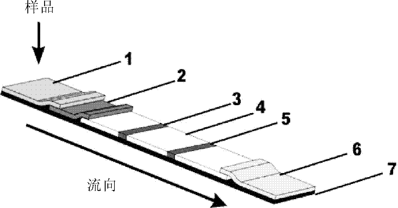
[0093] Embodiment 3 Preparation of immunochromatography detection reagent plate

[0094] (1) Preparation of antibody glass fiber strips labeled with gel-coated fluorescent microspheres

[0095] The D-dimer monoclonal antibody labeled with gel coated fluorescent latex particles prepared in Example 2 was mixed with 10 mM phosphate buffer solution containing 0.5% Triton X-100 (purchased from Sigma Company) and pH 7.4, and mixed to prepare 0.5 The solution with mg / ml concentration is evenly coated on glass cellulose paper, and the coating amount is 50μl / cm 2 , vacuum dried.

[0096] (2) Preparation of fluorescent latex-labeled rabbit IgG glass fiber strips

[0097] The gel-coated fluorescent latex-labeled rabbit IgG (purchased from Sigma) prepared by the same method as in Example 2 was mixed with 10 mM phosphate buffer solution containing 0.5% Triton X-100 and pH 7.4 to prepare 0.5 mg / The solution with a concentration of ml is evenly coated on glass cellulose paper, and the co...


PUM


Abstract

The invention provides a real-time quantitative detection reagent and a method of time-resolved fluorescence immune chromatography. The invention provides emulsion particles containing lanthanide elements coated by a gel, an antibody compound formed by the emulsion particles and an antibody coupled to the emulsion particles, a detection strip containing the antibody compound and a kit containing the detection strip, and applications thereof. The emulsion particles, the antibody compound and the kit can greatly eliminate non-specificity combination, and enables signal-to-noise ratio of detection signals to be increased significantly. The stability can be improved by coating the emulsion particles with the gel. The detection method has the advantages of high speed, high sensitivity, high specificity, etc.

Description

technical field [0001] The present invention relates to the field of detection. Specifically, the present invention relates to a real-time quantitative detection reagent and method of time-resolved fluorescence immunochromatography. Background technique [0002] Immunofluorescence technique (Immunofluorescence technique), also known as fluorescent antibody technology, is one of the earliest developed labeling immune techniques. It is a technique established on the basis of immunology, biochemistry and microscopy. Combine the antibody molecule with some tracer substances such as fluorescein, and after the antigen-antibody reaction, use a fluorescence microscope to judge the result. The main feature of this technology is strong specificity, but the quenching rate is high, unstable and difficult to preserve. [0003] Colloidal gold immunochromatography (GICA) technology is a simple and rapid immunological detection technology established on the basis of immunofiltration tech...

Claims


Application Information

Patent Timeline
IPC IPC(8): G01N33/533G01N33/543G01N33/53G01N33/531
Inventor 李小燕丁国荣
Owner 上海执诚生物科技有限公司
Why Eureka
Try Eureka