Microfluidic time-resolved fluorescence immunoassay device and application thereof

A time-resolved fluorescence and immunoassay technology, applied in the field of immunology, can solve the problems of high instrument use, high maintenance requirements, high comprehensive use cost, complex instrument structure, etc., and achieves improved experimental biological safety, simple structure, and simplified detection system. Effect

Active Publication Date: 2013-07-17
BEIJING BOHUI INNOVATION TECH
View PDF3 Cites 10 Cited by
  • Summary
  • Abstract
  • Description
  • Claims
  • Application Information

AI Technical Summary

Problems solved by technology

[0005] Fully automatic time-resolved fluorescence immunoassay analyzer means that the time-resolved fluorescence immunoassay (including adding samples, incubation, plate washing, adding enhancement solution, etc.) and fluorescence detection are all completed successively on the entire instrument, and almost no human participation is required; although It saves the tediousness and inaccuracy of manual sample addition, especially in the detection of a large number of s...

Method used


Image

  • Microfluidic time-resolved fluorescence immunoassay device and application thereof
  • Microfluidic time-resolved fluorescence immunoassay device and application thereof
  • Microfluidic time-resolved fluorescence immunoassay device and application thereof

Examples


Embodiment 1

[0058] Example 1: Microfluidic time-resolved fluorescent immunoassay device

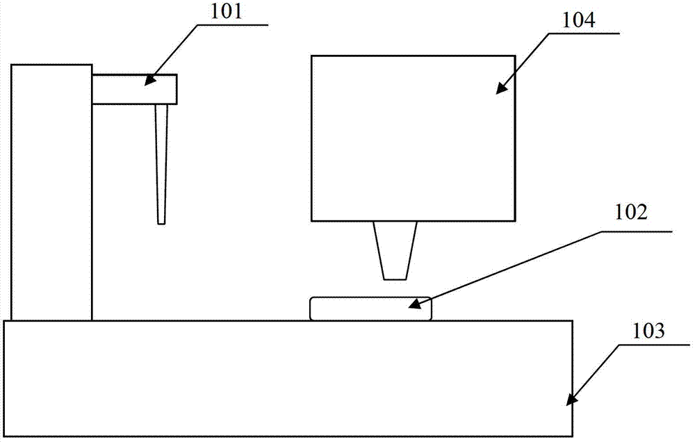
[0059] Such as figure 1 , figure 2 and image 3 As shown, a microfluidic time-resolved fluorescent immunoassay device includes a sample loading unit 101, a microfluidic control unit 103, a detection unit 104, and a microfluidic chip 102 driven by a membrane pump. The microfluidic chip 102 is located in the microfluidic chip holder on the analysis workbench of the microfluidic control unit 103 .

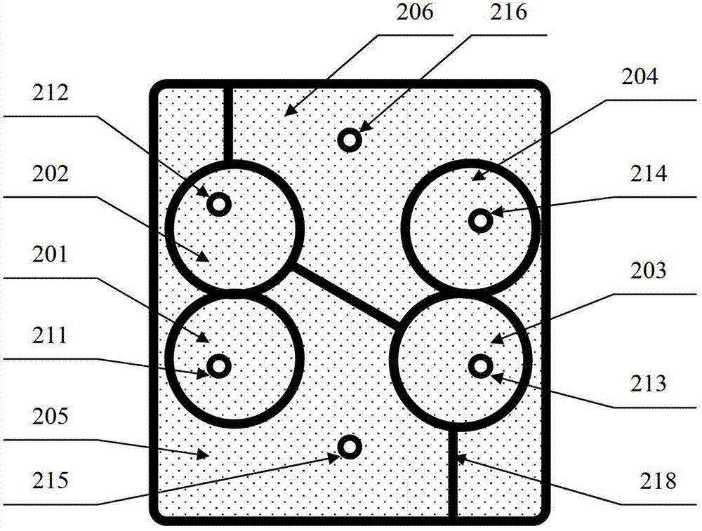
[0060] The microfluidic core 102 includes the following six pools: a sample pool 201, a diluent pool 202, a marker pool 203, a dissociation pool 204, a cleaning pool 205, and a waste pool 206, wherein the sample pool 201 is provided with a sample valve 301 and sample through-hole 211, diluent valve 302 and diluent through-hole 212 are provided in diluent pool 202, mark liquid valve 303 and mark liquid through-hole 213 are provided in mark liquid pool 203, dissociation liquid pool 204 is provided with D...

Embodiment 2

[0064] Embodiment 2: Measuring hepatitis B virus surface antigen with the device of embodiment 1

[0065] 1. Coating antibody: the antibody is anti-hepatitis B virus surface antigen (anti-HBs) monoclonal antibody

[0066] 1) Dilute the antibody 1:6000 times for coating with 50mM sodium carbonate-sodium bicarbonate buffer solution pH9.6, and set aside;

[0067] 2) Add the diluted antibody into the sample pool at 100 μl / well;

[0068] 3) The sample pool is placed in a 4°C environment and coated for 20 hours;

[0069] 4) After the coating is completed, wash 2 times with the washing liquid at 300 μl / well, and the washing liquid composition is 10 mM PBS at pH 7.4 containing 5% Tween-20 (volume ratio);

[0070] 5) After washing, place the sample pool on absorbent filter paper to absorb the remaining solution, then add blocking solution to the sample pool at an amount of 150 μl / well, and block for 2 hours at room temperature. 1%BSA (bovine serum albumin);

[0071] 6) After the se...

Embodiment 3

[0099] Embodiment 3: Measuring hepatitis B virus surface antigen with the device of embodiment 1

[0100] In step 4, the cleaning process is as follows: the one-way pump 405 works in one direction from the sample pool to the cleaning liquid pool, and sucks the cleaning liquid into the sample pool 201 .

[0101] With the diluent pool 202 as the cleaning buffer pool, the sample pool 201 first gets rid of the waste liquid, sucks the cleaning liquid, then closes the one-way pump sample cleaning pump 405 and the one-way pump sample waste liquid pump 406; starts the two-way pump 402 to make the cleaning liquid in Flow between the sample pool 201 and the diluent pool 202 for cleaning; the cleaning liquid is in the sample pool 201 when stopped.

[0102] Other steps are the same as in Example 2.


PUM


Abstract

The invention provides a microfluidic time-resolved fluorescence immunoassay device. The device comprises a sample injection unit (101), a microfluidic control unit (103), a detection unit (104) and a microfluidic chip (102) adopting a membrane pump drive mode, and the microfluidic chip (102) is positioned in a microfluidic chip carrier on an analysis workbench of the microfluidic control unit (103). The microfluidic time-resolved fluorescence immunoassay device has the advantages that by adopting the microfluidic technology, a detection system is simplified; and a microplate oscillator and a microplate washer are omitted, a microfluidic control system is simple in structure, optimized, free of mechanical movements and structures of cleaning, vibration, transmission and the like, and automation is achieved conveniently.

Description

technical field [0001] The invention belongs to the field of immunology, in particular to a time-resolved fluorescence analysis device and its application. Background technique [0002] Time-resolved fluorescence analysis technology is a new type of non-radioactive micro-analysis technology based on lanthanide elements with unique fluorescence characteristics and their chelating agents as tracers. The rapid development of TRFIA methodological research and clinical application has become a new milestone in the development of marker immunoassay. TRFIA technology has the advantages of high sensitivity, wide range of standard curve, easy operation, no radioactive pollution, and multiple labels. It is widely used in tumors, The clinical laboratory diagnosis of infectious diseases, endocrine diseases, autoimmune diseases, and genetic diseases has become one of the commonly used analysis methods in biomedical research and clinical ultra-trace biochemical tests. [0003] TRFIA (Tim...

Claims


Application Information

Patent Timeline
IPC IPC(8): G01N21/64G01N33/533B01L3/00
Inventor 杨奇
Owner BEIJING BOHUI INNOVATION TECH
Why Eureka
Try Eureka