

Acellular biological patch, preparation method and apparatus thereof
A biological patch and decellularization technology, applied in the field of natural decellularized biological patch and its preparation, can solve the problems of insufficient decellularization and antigen removal, inflammatory reaction, immune rejection, etc., to overcome uneven mechanical strength, Avoid immune rejection and be beneficial to industrialized production
- Summary
- Abstract
- Description
- Claims
- Application Information
AI Technical Summary
Problems solved by technology
Method used
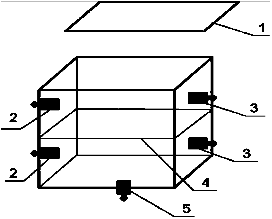
Image

Examples
Embodiment 1
[0055] Embodiment 1, One of the preparation method and device of decellularized biological patch
[0056] The preparation method and steps of the present embodiment are as follows:
[0057] Step 1. Pretreatment: Wash the pig small intestine that has been cut open with the mucous membrane layer and lymph nodes removed with purified water until there is no dirt in the intestine, then soak it in aqueous hydrochloric acid solution with a volume concentration of 2.0% for 2.5 hours, and then use purified Wash with water until the pH value is 6.0-8.0.
[0058] Step 2. Disinfection treatment: Place the pig small intestine treated in step 1 in a hydrogen peroxide solution with a volume concentration of 15%, soak for 0.5 hours, wash with purified water for 3 times, scrape off the muscular layer and serosal layer with a blade, and retain the mucous membrane Lower layer: Soak the small intestinal submucosa in 70% ethanol solution for 1 hour, then rinse with purified water until odorless....
Embodiment 2
[0065] Embodiment 2, the preparation method and device of decellularized biological patch 2
[0066] The preparation method and steps of the present embodiment are as follows:
[0067] Step 1. Pretreatment: Wash the pig small intestine that has been dissected by removing the mucous membrane layer and lymph nodes with purified water until there is no dirt in the intestine, then soak it in an aqueous solution of lactic acid with a volume concentration of 3.0% for 1.5 hours, and then use purified Wash with water until the pH value is 6.0-8.0.
[0068] Step 2. Disinfection treatment: place the pig small intestine treated in step 1 in a hydrogen peroxide solution with a volume concentration of 1%, soak for 2.5 hours, wash with purified water twice, scrape off the muscular layer and serosa layer with bamboo slices, and keep Submucosa: After soaking the submucosa of the small intestine in isopropanol solution for 3 hours, rinse it with purified water until it is odorless.
[0069] ...
Embodiment 3
[0073] Embodiment 3, the third preparation method and device of decellularized biological patch
[0074] The preparation method and steps of the present embodiment are as follows:
[0075] Step 1, pre-treatment: the pig small intestine that has been removed from the mucous membrane layer and lymph nodes is washed with purified water until there is no dirt in the intestine, and then soaked in a peracetic acid solution with a volume concentration of 1.5% for 1.0 hour, and then Wash with purified water to pH 6.0-8.0.
[0076] Step 2. Disinfection treatment: place the pig small intestine treated in step 1 in 3% hydrogen peroxide solution, soak for 1.5 hours, wash with purified water for 3 times, scrape off the muscular layer and serosal layer with a stainless steel plate, and keep Submucosa: Soak the submucosa of the small intestine in 80% ethanol solution for 2 hours, then rinse with purified water until odorless.
[0077] Step 3, decellularization and antigen removal: using a ...
PUM
 Abstract
Description
Claims
Application Information