Indolpyridine type fluorescent probe for imaging RNA and nucleolus in living cell

A fluorescent probe, indolepyridine technology, applied in the field of fluorescent probes, can solve the problems of unclear mechanism of interaction between small molecules and RNA, limitations of pathology research and drug development, difficulties in RNA probe research, etc., and achieve photostability Strong, good membrane permeability, strong color effect

Active Publication Date: 2013-08-28
SHANDONG UNIV
View PDF4 Cites 23 Cited by
  • Summary
  • Abstract
  • Description
  • Claims
  • Application Information

AI Technical Summary

Problems solved by technology

Compared with many commercial DNA probes, there are very few RNA probes, because the existing nucleic acid probes generally have greater affinity with double-stranded DNA, and the mechanism of action between small molecules and RNA is not yet clear, so RNA probes research is more diffic...

Method used


Image

  • Indolpyridine type fluorescent probe for imaging RNA and nucleolus in living cell
  • Indolpyridine type fluorescent probe for imaging RNA and nucleolus in living cell
  • Indolpyridine type fluorescent probe for imaging RNA and nucleolus in living cell

Examples


Embodiment 1

[0026] Example 1: Synthesis of (E)-4-(2-(1H-indole-3-)vinyl)-1-picoline iodide salt (2c)

[0027] Dissolve N-methyl-2-formylpyrrole and N-hydroxyethyl-4-picoline in methanol to obtain a light yellow transparent solution, add 3-4 drops of piperidine, and the solution turns red rapidly. After reflux reaction for 4h, a purple-red precipitate precipitated out. Cool, filter, and wash with dichloromethane to obtain a brown powder with a yield of 90%.

[0028] 1 H NMR (300MHz, DMSO-d 6 ),δ(ppm):11.92(s,1H),8.68(d,J=7.0Hz,2H),8.24(d,J=16.2Hz,1H),8.16(dd,J=6.6,1.7Hz,1H ),8.11(d,J=6.92Hz,2H),7.97(s,1H),7.51(dd,J=6.8,1.7Hz,1H),7.26(m,3H),4.18(s,3H).13C NMR (400MHz, DMSO-d6), δ (ppm): 154.62, 144.62, 137.96, 136.69, 132.71, 125.36, 123.38, 122.08, 121.59, 120.90, 117.29, 114.02, 113.05, 46.74. HRMS (m / z): [ M-I]+. Calcd for C16H15IN2, 235.12; found, 235.12.

Embodiment 2

[0029] Example 2: HeLa, SiHa and H17702 cell culture

[0030] HeLa, SiHa and H17702 cells were adherently cultured in culture medium containing 10% fetal bovine serum at 37°C, 5% CO 2 Cultured in a saturated humidity incubator, and subcultured once every 2-3 days.

[0031] When the cells grow to the logarithmic phase, culture the slices: ① Soak the coverslips in absolute ethanol for 30 minutes, dry them with an alcohol lamp and place them in a disposable 35mm culture dish for later use; Wash the cells three times with PBS, digest with 1 mL of 0.25% trypsin for 3-5 minutes, pour out the trypsin carefully, add fresh culture medium and pipette evenly, and count the cells. Concentration is 1x10 5 , and then inoculated into the above-mentioned petri dish with coverslip in 5% CO 2 Cultured in an incubator to allow the cells to grow on the sheet.

[0032] After the cell slides grow and cover the coverslip, they are used for experiments.

Embodiment 3

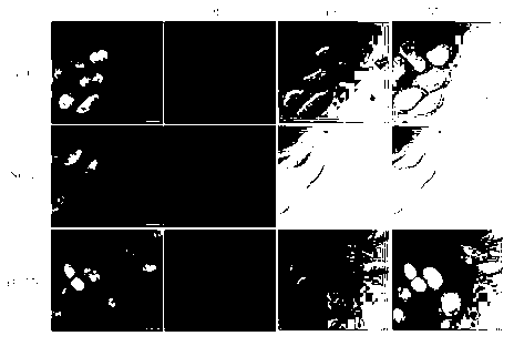
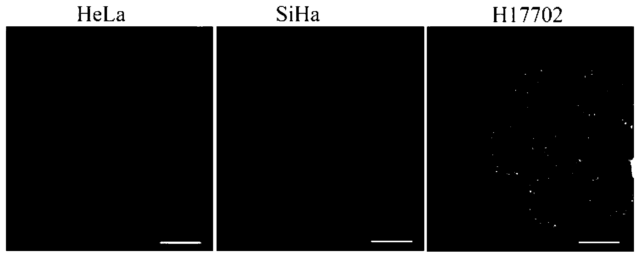
[0033] Example 3: (E)-4-(2-(1H-indole-3-)vinyl)-1-picoline iodide salt (2c) staining observation on HeLa, SiHa and H17702 cells

[0034] Wash the coverslips (climbing slides) covered with HeLa, SiHa and H17702 cells prepared in Example 2 three times with PBS, and then use (E)-4-(2- (1H-indole-3-)vinyl)-1-picoline iodide fluorescent probe solution in CO 2 In the incubator, stain the various cells for 30 min respectively.

[0035] Take out the stained slides, wash away the unbound excess dye solution, cover the cell growth side down on the glass slide, observe under the fluorescence microscope and laser scanning confocal lens, and record the stained parts, fluorescence distribution and brightness in the cells changes etc.

[0036] see results figure 1 , figure 2 . The fluorescence image shows obvious green light distribution in the cytoplasm and nucleolus, clearly suggesting that the probe of the present invention can specifically image RNA in the cytoplasm and nucleolus i...


PUM


Abstract

The invention discloses an indolpyridine type fluorescent probe for imaging RNA (Ribonucleic Acid) and nucleolus in a living cell. The structural general formula of the fluorescent probe is shown in the specification, wherein R1 and R2 represent alkyl, hydroxyalkyl or ether group, respectively. The invention also discloses application of the fluorescent probe in marking or displaying of the distribution of the RNA and the nucleolus in the living cell and distinguishing of the dead state and the live state of the cell. The probe provided by the invention has the characteristics of wide application range, good membrane permeability, low cytotoxicity and ability of exclusively realizing fluorescent imaging of RNA in the living cell.

Description

technical field [0001] The invention relates to a fluorescent probe, in particular to an indolepyridine salt fluorescent probe used for imaging RNA and nucleoli in living cells. Background technique [0002] Nucleic acid is not only the basic component of all biological cells, but also plays a dominant role in the growth, development, reproduction, inheritance and variation of organisms and other major life phenomena. For example, the oxidative decomposition product of nucleic acid - purine is the main cause of human uric acid increase and gout. Nucleic acid macromolecules are divided into two categories: deoxyribonucleic acid (DNA) and ribonucleic acid (RNA), which play a role in storing and transmitting genetic information in the replication and synthesis of proteins. DNA is the main material basis for storing, replicating and transmitting genetic information. RNA plays an important role in the process of protein synthesis, in which transfer RNA (tRNA) carries and transfe...

Claims


Application Information

Patent Timeline
IPC IPC(8): C09K11/06C07D401/06G01N21/64
Inventor 于晓强孙渝明宋国芬
Owner SHANDONG UNIV
Why Eureka
Try Eureka