Protein covalent coupling method on surface of magnetic beads

A surface covalent, protein-based technology, applied in the direction of material inspection products, measuring devices, instruments, etc., can solve the problems of self-agglutination of coupled magnetic beads, low coupling efficiency of magnetic beads, non-specific adsorption of magnetic beads, etc., and achieve protein coupling High coupling efficiency, increased coupling efficiency, and good repeatability

Inactive Publication Date: 2013-09-25
CAPITALBIO CORP +1
View PDF5 Cites 15 Cited by
  • Summary
  • Abstract
  • Description
  • Claims
  • Application Information

AI Technical Summary

Problems solved by technology

Due to the difference in the choice of activator, activation conditions, coupling conditions and coupling process, the magnetic beads coupled with protein have low coupling e

Method used


Image

  • Protein covalent coupling method on surface of magnetic beads
  • Protein covalent coupling method on surface of magnetic beads
  • Protein covalent coupling method on surface of magnetic beads

Examples


Embodiment 1

[0032] Example 1, carboxyl magnetic beads coupled goat anti-fluorescein isothiocyanate antibody

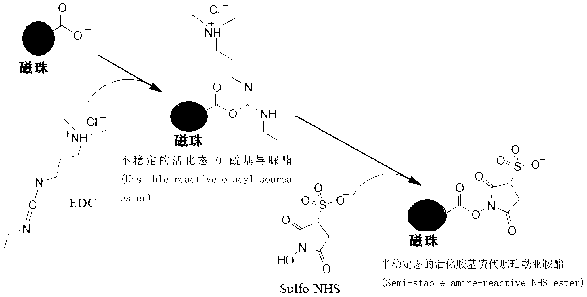
[0033] 1. Activation of carboxyl magnetic beads

[0034] The purpose of this step is to generate carbodiamine-containing chemical groups on the surface of the magnetic beads.

[0035] 1. Magnetic bead pretreatment

[0036] Take 1000 mg carboxy magnetic bead colloidal solution and settle under the action of magnetic field for 20 minutes, discard the supernatant, and then wash the magnetic beads once with 50 ml pH7.0, 0.01M MES buffer.

[0037] 2. Fully suspend the magnetic beads obtained in step 1 with 20ml pH7.0, 0.01M MES buffer solution, then add EDC and NHS, so that the concentration of EDC is 0.1mM, and the concentration of NHS is 100mM, shake the reaction at room temperature (30 rpm / min) for 240 minutes to obtain an activated magnetic bead solution.

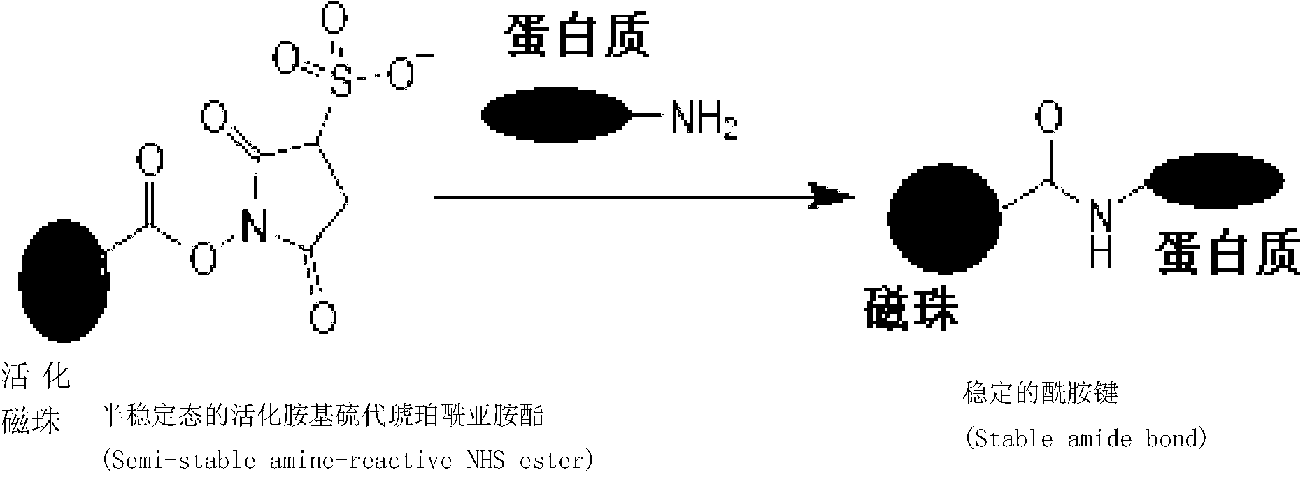
[0038] 2. Goat anti-fluorescein isothiocyanate antibody coupled to carboxyl magnetic beads

[0039] 1. Take 10ml goat ant...

Embodiment 2

[0066] Example 2, carboxyl magnetic beads coupled monoclonal antibody

[0067] 1. Activation of carboxyl magnetic beads

[0068] 1. Magnetic bead pretreatment

[0069] Take 200 mg carboxyl magnetic bead colloidal solution and settle under the action of a magnetic field for 10 minutes, discard the supernatant, and wash the sedimented magnetic beads with pH 4.0, 0.1 M MES buffer 3 times, 20 ml each time.

[0070] 2. Fully suspend the magnetic beads obtained in step 1 with 20ml pH4.0, 0.1M MES buffer, then add EDC and NHS, make the concentration of EDC 30mM, and the concentration of NHS 0.3mM, shake the reaction at room temperature (20 rpm / min) for 10 minutes, which is the activated magnetic bead solution.

[0071] 2. Carboxyl magnetic beads conjugated monoclonal antibody

[0072] 1. Take thyroxine monoclonal antibody, load it on a Sephadex G25 chromatographic column, elute with pH 4.0, 0.1M MES buffer, and collect 4ml of antibody solution.

[0073] 2. Coupling

[0074] Add...


PUM


Abstract

The invention discloses a protein covalent coupling method on the surface of magnetic beads. The method comprises the steps that: (1) the magnetic beads and an activating agent are subjected to co-incubation, such that activated magnetic beads are obtained; (2) the activated beads are subjected to co-incubation with protein, such that magnetic beads coupled with the protein are obtained; (3) the magnetic beads coupled with the protein are treated by using a blocking solution, such that surface active sites are blocked; and (4) the magnetic beads obtained in the step (3) are subjected to simultaneous vibration and sieving, and the sieved magnetic beads are collected. The method provided by the invention has the advantages of high protein coupling efficiency, good repeatability, and versatility. With the method, biological activities of protein coupled on the magnetic beads are not influenced. With the method provided by the invention, the obtained magnetic beads with covalently coupled protein can be used in fields such as immunoassay, biochemical detection, molecular detection, cell typing, and the like.

Description

technical field [0001] The invention relates to a method for covalently coupling proteins on the surface of magnetic beads. Background technique [0002] Magnetic beads, also known as magnetic particles, are a kind of spherical composite materials with a diameter of nanometers or micrometers. Magnetic beads are composed of three parts, the core is a small metal particle (iron oxide, ferric oxide), a layer of polymer material (such as polystyrene, polyvinyl chloride, etc.) is evenly wrapped around the core, and the outermost layer is a functional base layer. With functional groups such as amino (-NH 2 ), carboxyl (-COOH), hydroxyl (-OH), etc. [0003] The magnetic beads are uniform spherical and have superparamagnetism, so that the magnetic beads can move in the direction of the magnetic field under the action of an external magnetic field to achieve the purpose of separating the target. The chemical functional groups on the outermost layer are used to bind with biologicall...

Claims


Application Information

Patent Timeline
IPC IPC(8): G01N33/68G01N33/531G01N33/543
Inventor 郭建夫杨宝君韩学栋周玉祥
Owner CAPITALBIO CORP
Why Eureka
Try Eureka