Suspending skin tissue engineering nanofiber support and preparation method thereof

A skin tissue engineering and nanofiber technology, applied in the field of suspendable skin tissue engineering nanofiber scaffolds and its preparation, can solve the problems of unfavorable cell adhesion and specific gene activation, the risk of infecting certain diseases, and the lack of cell recognition signals , to achieve good encapsulation and film-forming properties, to achieve flatness and integrity, high strength and hardness

Active Publication Date: 2015-01-14
常熟紫金知识产权服务有限公司
View PDF6 Cites 0 Cited by
  • Summary
  • Abstract
  • Description
  • Claims
  • Application Information

AI Technical Summary

Problems solved by technology

Scaffold materials for biological skin tissue engineering mainly include collagen and acellular dermal matrix. The former is a classic artificial skin scaffold material, but its disadvantage is that it has poor mechanical properties and is not easy to operate. Decellularized cadaver skin or pigskin is used as the scaffold material to prepare artificial skin. Its advantages are good mechanical properties and low antigenicity. The disadvantage is that the material source is very limited and there is a risk of infecting certain diseases, which limits its application.
Synthetic skin tissue engineering scaffold materials are mainly polylactide, polylactone, polyhydroxyalkanoate, and polycarbonate, but their biggest disadvantage is the lack of cell recognition signals, which is not conducive to cell adhesion and activation of specific genes

Method used


Image

  • Suspending skin tissue engineering nanofiber support and preparation method thereof
  • Suspending skin tissue engineering nanofiber support and preparation method thereof

Examples


Embodiment 1

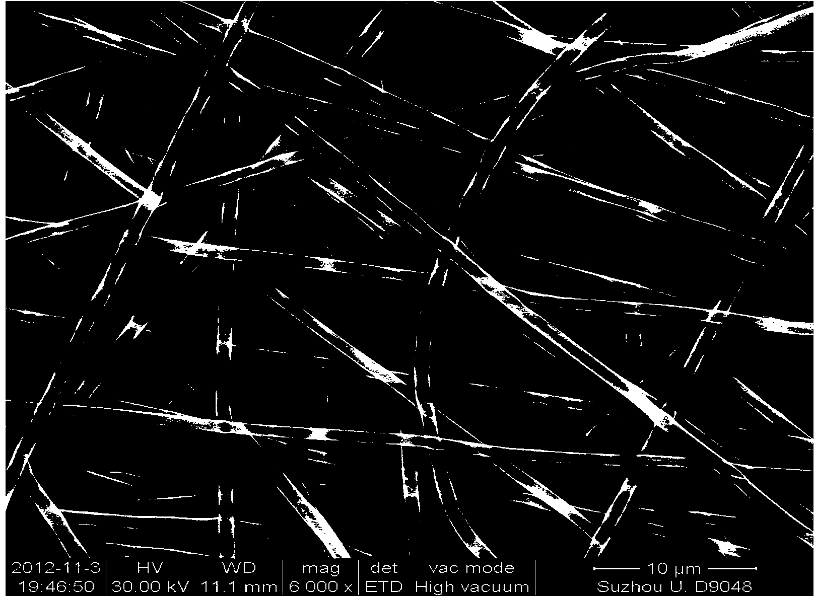
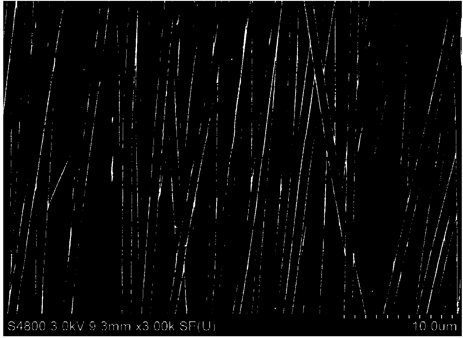
[0042] First, a polylactic acid-glycolic acid copolymer solution with a concentration of 0.3 g / ml is prepared, and the solvent is a tetrahydrofuran / acetic acid mixture with a volume ratio of 4:1. The solution is placed at room temperature for 24 hours, and nanofiber membranes are prepared by electrostatic spinning after the solute is uniformly dissolved. Electrospinning process parameters: voltage is 6000V; electrode spacing is 10cm; solution supply flow is 40μl / h; receiving shaft diameter is 20mm; receiving speed is 2000r / min; receiving time is 1.5h. After the electrostatic spinning is finished, the receiving device is grounded. After the electrostatic charge is eliminated, the nanofiber membrane is removed from the receiving shaft, and the resulting nanofiber membrane is cut into the desired shape. The fiber membrane and the auxiliary scaffold are bonded together to finally obtain the nanofiber scaffold that can suspend skin tissue engineering.

Embodiment 2

[0044] First, a polylactic acid-glycolic acid copolymer solution with a concentration of 0.4 g / ml is prepared, and the solvent is a tetrahydrofuran / acetic acid mixture with a volume ratio of 4:1. The solution is placed at room temperature for 24 hours, and nanofiber membranes are prepared by electrostatic spinning after the solute is uniformly dissolved. Electrospinning process parameters: voltage is 4000V; electrode spacing is 10cm; solution supply flow is 20μl / h; receiving shaft diameter is 20mm; receiving speed is 2000r / min; receiving time is 2h. After the electrostatic spinning is finished, the receiving device is grounded. After the electrostatic charge is eliminated, the nanofiber membrane is removed from the receiving shaft, and the resulting nanofiber membrane is cut into the desired shape. The fiber membrane and the auxiliary scaffold are bonded together to finally obtain the nanofiber scaffold that can suspend skin tissue engineering.

Embodiment 3

[0046] First, a polylactic acid-glycolic acid copolymer solution with a concentration of 0.2g / ml is prepared, and the solvent is a tetrahydrofuran / acetic acid mixture with a volume ratio of 4:1. The solution is placed at room temperature for 24 hours, and nanofiber membranes are prepared by electrostatic spinning after the solute is uniformly dissolved. Electrospinning process parameters: voltage is 10000V; electrode spacing is 20cm; solution supply flow rate is 60μl / h; receiving shaft diameter is 20mm; receiving speed is 2000r / min; receiving time is 1h. After the electrostatic spinning is finished, the receiving device is grounded. After the electrostatic charge is eliminated, the nanofiber membrane is removed from the receiving shaft, and the resulting nanofiber membrane is cut into the desired shape. The fiber membrane and the auxiliary scaffold are bonded together to finally obtain the nanofiber scaffold that can suspend skin tissue engineering.


PUM

PropertyMeasurementUnit
concentrationaaaaaaaaaa

Abstract

The invention discloses a suspending skin tissue engineering nanofiber support and a preparation method of the suspending skin tissue engineering nanofiber support. The support is composed of a polylactic acid-glycolic acid copolymer nanofiber membrane and a polypropylene auxiliary strut, wherein the nanofiber membrane is prepared through an electrospinning technology, through control over process parameters and using of different receiving devices, different structures with orderly or unordered fiber arrays are obtained, and the structures are adhered together through heating and the auxiliary strut. The obtained nanofiber support can suspend on the surface of a culture solution, keep straight performance and integrity for a long time and simulate the growing environment of skin cells and is beneficial to adherency, growth, migration, multiplication and differentiation of the skin cells, and finally the tissue engineering dermis is formed. The suspending skin tissue engineering nanofiber support and the preparation method are simple in preparation technology, low in cost and easy to apply widely.

Description

Technical field [0001] The invention relates to the technical fields of tissue engineering and micro-nano manufacturing, in particular to a suspendable skin tissue engineering nanofiber scaffold and a preparation method thereof. Background technique [0002] The skin is one of the important organs of the human body and can be completely renewed once within 60 days. The skin not only protects the human body from dirt or bacteria, but also keeps the moisture in the human body from being lost. When a large area of ​​skin is severely burned or damaged, the doctor must immediately infuse the fluid and protect the wound. If only the superficial layer of the skin is damaged, the new skin will regenerate. If the patient suffers a severe burn, the skin cannot be repaired by itself. For a long time, people can only repair the wounds with severe skin defects by cutting the skin grafts from normal parts of the body. Although it can heal the wounds, it leaves new wounds at the skin removal ...

Claims


Application Information

Patent Timeline
Patent Type & Authority Patents(China)
IPC IPC(8): A61L27/18A61L27/16A61L27/60D04H1/435
Inventor 汝长海王飞龙陈瑞华杨湛
Owner 常熟紫金知识产权服务有限公司
Why Eureka
Try Eureka