Inflammasome activity reporting system for sub-cellular localization and application thereof

An inflammasome and activity detection technology, applied in the field of biomedicine, can solve the problems of cumbersome operation, small protein molecular weight, and difficulty in obtaining stable results, and achieve high activation efficiency

Inactive Publication Date: 2014-09-03
SUN YAT SEN UNIV
View PDF1 Cites 7 Cited by
  • Summary
  • Abstract
  • Description
  • Claims
  • Application Information

AI Technical Summary

Problems solved by technology

At present, the detection of inflammasome activity mainly relies on ELISA and Western Blot technology to detect the shearing of mature IL-1β and Caspase-1 respectively, and the operation is cumbersome
And because the molecular weight of the corresponding protein is small (mature IL-1β is 17Kda, while the P20 sub...

Method used


Image

  • Inflammasome activity reporting system for sub-cellular localization and application thereof
  • Inflammasome activity reporting system for sub-cellular localization and application thereof
  • Inflammasome activity reporting system for sub-cellular localization and application thereof

Examples


Embodiment 1

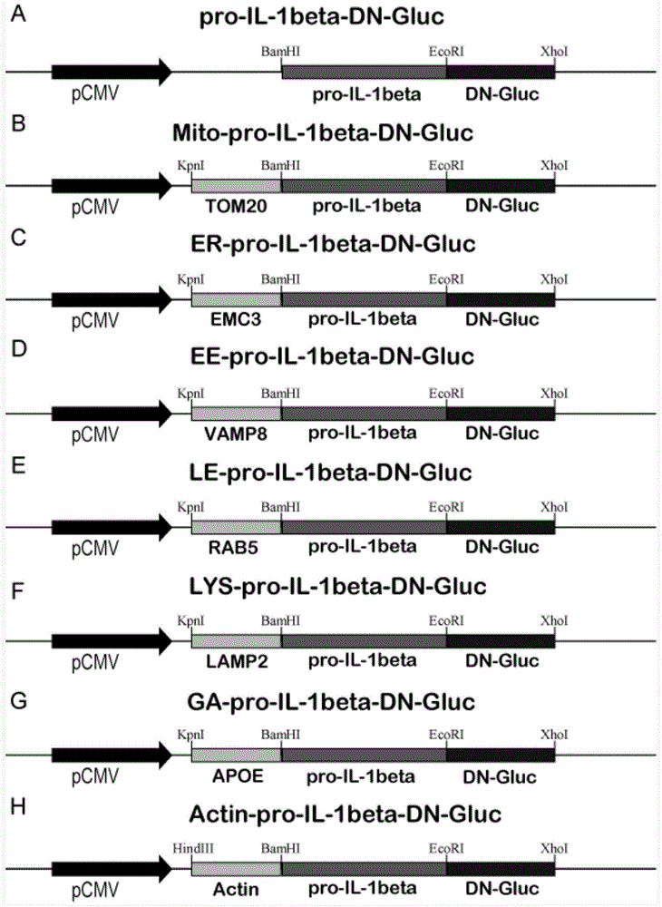
[0046] Method for constructing recombinant gene (method for constructing subcellular localized inflammasome reporter gene / plasmid)

[0047] (1) Construction of the pro-IL-1β-DN-Gluc recombinant plasmid:

[0048] (1) Acquisition of DN-Gluc gene: design primers according to the sequence of DN-Gluc gene, use the Actin-DN-Gluc plasmid [KETTELER, R., Z.SUN, et al.A pathway sensor for genome provided by Ketteler et al. -wide screens of intracellular proteolytic cleavage.Genome Biol,2008,9(4):R64.] as a template, using an upstream primer (SEQ ID No.17) 5'-TACGGGAATTCATGCTAGCCAAGCCCAC-3' with an EcoRI restriction site and The downstream primer (SEQ ID No.18) 5'-TACGCAGATCTAGACATGATAAGATAC-3' with XhoI restriction site was used for PCR to amplify and obtain DN-Gluc; perform PCR reaction according to PrimerStar2×PCR Mix (Bao Biological Engineering, Dalian): The total PCR reaction system was 50 μL, which contained 25 μL of PrimerStar PCR Mix, 0.2 μmol / L of upstream and downstream primer...

Embodiment 2

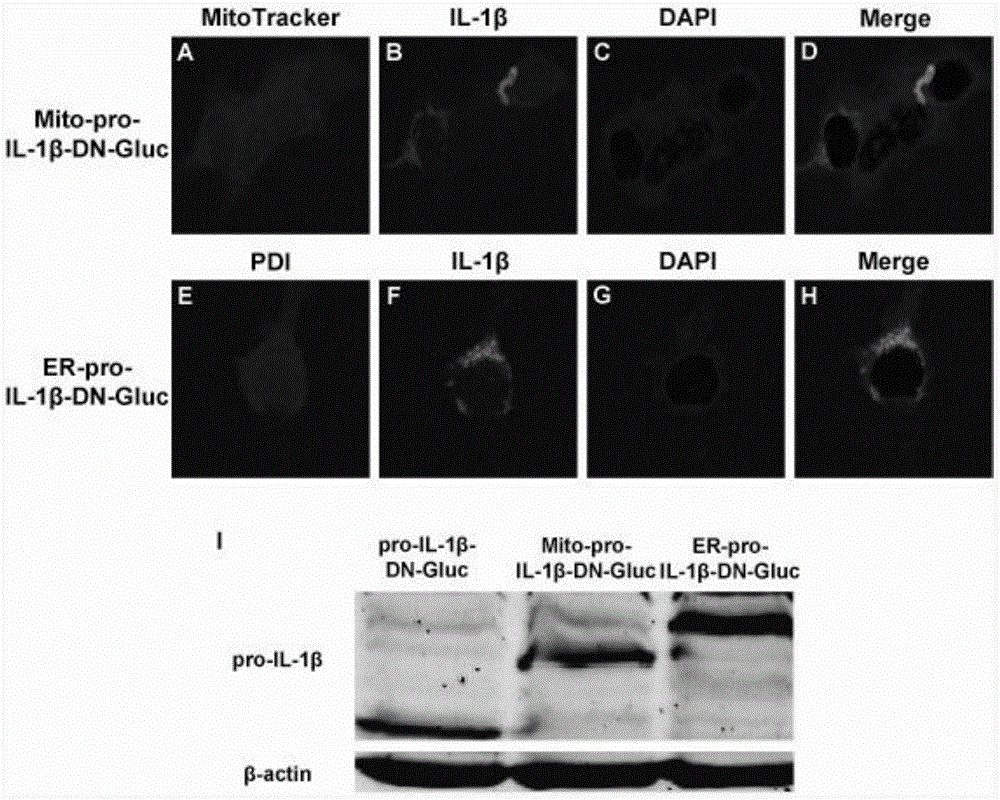
[0073] Expression and localization detection of fusion protein

[0074] (1) Plasmid transfection: Inoculate 293T into 12-well plates with cell slides at a density of 4×10 5 / cm 2 . After 24 hours, 0.8 μg of plasmid was transfected per well, and the transfection reagent was Lipofectamine2000 (Invitrogene Company). After 24 hours, the cells were harvested for immunofluorescence staining and Western Blot.

[0075] (2) Immunofluorescence staining and confocal laser microscope observation: the mitochondrial staining group was added with MitoTracker and cultured for 2 hours. Wash twice with PBS; add fixative solution and incubate at room temperature for 10 minutes; wash twice with PBS; add perforation solution and incubate at room temperature for 10 minutes; add blocking solution and incubate at room temperature for 1 hour; wash twice with PBS; Corresponding solution (PBS solution with V / V of 0.1% BSA), incubate at room temperature for 1 h; wash with PBS 3 times; secondary antib...

Embodiment 3

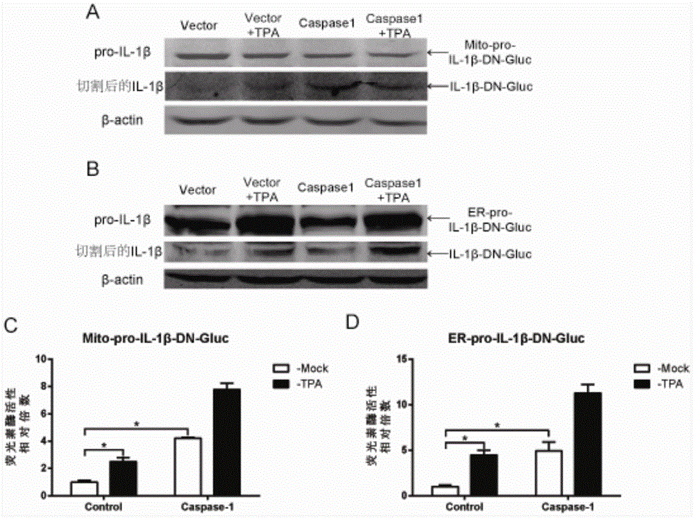
[0078] Responses of different detection reagents / reporter systems to inflammasome activation induced by TPA stimulation and transfection of Caspase-1

[0079] (1) Plasmid transfection: respectively inoculate 293T into 12-well plates at a density of 4×10 5 / cm 2 . After 24 hours, each well was transfected with 0.4 μg reporter plasmid and pCMV-3myc-Caspase-120ng, and the control group was co-transfected with pCMV-3myc20ng, and the transfection reagent was Lipofectamine2000 (Invitrogene Company). After 30 hours, the cell supernatant was collected to detect luciferase activity, and the cells were collected for Western Blot.

[0080] (2) Cell stimulation: 24 hours after transfection, the cell culture medium was discarded and replaced with fresh DMEM (serum-free, containing 100 ng / mL TPA), and the control group was replaced with fresh DMEM without TPA and serum.

[0081] (3) Western Blot is the same as in Example 2. The results showed that both TPA and transfection of Caspase-1 ...


PUM


Abstract

The invention belongs to the field of biomedicine, and relates to a fusion protein for sub-cellular localization and an application thereof. A system can be used for reporting the activity of inflammasomes on the organelle level mainly based on secretion type luciferase. 7 kinds of plasmids included in the system are mainly obtained by sequentially cloning an organelle localization gene, an interleukin 1beta precursor coding gene (pro-IL-1beta) and secretion type luciferase (DN-Gluc) to a multiple cloning site zone of an expression vector pcDNA3.1. The plasmids included in the system are transfected to target cells respectively, and the activity of the inflammasomes can be quantitatively analyzed by detecting the activity of luciferase in extracellular supernate, so that the system has the advantages of high efficiency, fastness, simplicity, convenience and the like and can be applied to medicament screening, molecular mechanisms, live animal research and development of related products related to the inflammasomes.

Description

technical field [0001] The invention belongs to the field of biomedicine, and relates to a fusion protein, its coding sequence and application. Background technique [0002] Inflammasomes (Inflammasomes) mediate the body's anti-infection immunity and are an important part of innate immunity. They mainly process interleukin-1β (Interleukin-1β, IL-1β) and interleukin-18 precursors through Caspase-1 to produce corresponding The mature cytokines regulate inflammation and cell death (Pyroptosis). The inflammasome has been shown to be involved in various diseases and cancers. At present, the detection of inflammasome activity mainly relies on ELISA and Western Blot technology to detect the shearing of mature IL-1β and Caspase-1 respectively, and the operation is cumbersome. And because the molecular weight of the corresponding protein is small (mature IL-1β is 17Kda, and the P20 subunit of Caspase-1 is 20Kda), and has strong extracellular secretion ability, it is often difficult...

Claims


Application Information

Patent Timeline
IPC IPC(8): C12N15/62C12N9/02C12Q1/66C12Q1/37
Inventor 况二胜李宇清
Owner SUN YAT SEN UNIV
Why Eureka
Try Eureka