Eureka AIR delivers breakthrough ideas for toughest innovation challenges, trusted by R&D personnel around the world.

Adipose-derived stem cell transfected by magnetic nanoparticle mediated IGF-I gene and preparation method thereof

A magnetic nanoparticle and adipose stem cell technology, which can be applied to cells modified by the introduction of foreign genetic material, introduction of foreign genetic material using a carrier, recombinant DNA technology, etc. , target cells and cytokines cannot aggregate, proliferate and other problems, to achieve the effect of high targeting and improving gene transfection efficiency

Inactive Publication Date: 2015-05-20
HANGZHOU CITY XIAOSHAN DISTRICT TRADITIONAL CHINESE MEDICAL HOSPITAL
View PDF0 Cites 3 Cited by
  • Summary
  • Abstract
  • Description
  • Claims
  • Application Information

AI Technical Summary

Problems solved by technology

[0010] When the composite scaffold loaded with IGF-I and seed cells is implanted in the animal model, due to the extremely low homing rate of stem cells, coupled with the daily activities of the animal model and the flow of blood and joint fluid at the scaffold in the model, The target cells and cytokines loaded in the scaffold cannot effectively gather and proliferate for a long time, which seriously affects the normal repair speed and degree of defective cartilage

Method used


Image

  • Adipose-derived stem cell transfected by magnetic nanoparticle mediated IGF-I gene and preparation method thereof
  • Adipose-derived stem cell transfected by magnetic nanoparticle mediated IGF-I gene and preparation method thereof
  • Adipose-derived stem cell transfected by magnetic nanoparticle mediated IGF-I gene and preparation method thereof

Examples


Embodiment 1

[0031] Example 1 Isolation and culture of rabbit ADSCs

[0032] After ether anesthesia, the adipose tissue in the groin of the rabbit was taken under aseptic conditions, blood vessels and other tissues were removed, and the PBS buffer containing double antibodies was repeatedly washed to remove impurities and blood cells. After the adipose tissue was fully cut with scissors, it was placed in 0.2% Type collagenase, 37 ° C shaker 100r / min digestion 40min; digestion with an equal volume of DMEM medium containing 10% fetal bovine serum to stop. The reaction solution was centrifuged at 800r / min for 10min, and the fat and supernatant in the upper layer were discarded; the sediment was made into a suspension in DMEM medium with 10% fetal bovine serum, and filtered with a 100um sieve to remove cell clumps. Cells were seeded in 25 cm culture flasks at 37°C, 5% CO 2 Routine culture in the incubator, change the medium after 24h, remove non-adherent cells and remaining blood cells. ...

Embodiment 2

[0033] Example 2 Iron oxide magnetic nanoparticles (Fe 3 O 4 MNPs) preparation and detection

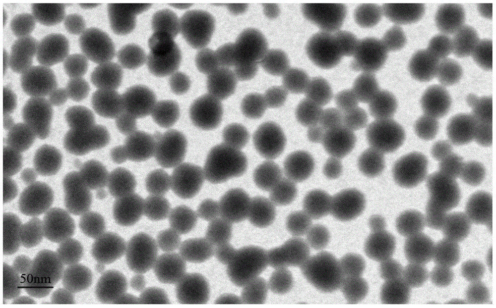
[0034] 1. Fe 3 O 4 Preparation of MNPs

[0035] Add 100 mL of 2-pyrrolidone and 10 mmol of acetylacetone into a 250 mL 3-necked bottle, remove the air, stir magnetically under the protection of argon, and boil and reflux at 245 °C for 30 min. Heating was stopped, and the argon continued to protect and cool to room temperature. A large amount of methanol was quickly added to obtain a black precipitated ferrofluid. After washing with acetone three times, MNPs were obtained after vacuum drying at 60°C, and stored in a desiccating dish.

[0036] , Fe 3 O 4 Characterization analysis and morphological observation of MNPs

[0037] The phase analysis of MNPs was carried out by X-ray diffraction (XRD). Fe was diluted with PBS at pH 7.0 3 o 4 The MNPs sample was ultrasonically oscillated at 200W for 3 min, and the sample was dropped on a copper grid. Af...


PUM


Abstract

The invention relates to the field of articular cartilage injury repair and particularly relates to an adipose-derived stem cell transfected by a magnetic nanoparticle mediated IGF-I gene and a preparation method thereof. The method comprises three steps of ADSCs separation and culture, Fe3O4 MNPs preparation and in vitro transfection. The mediated IGF-I gene with an iron oxide magnetic nanoparticle as a vector is used for transfecting ADSCs to achieve over expression of IGF-I, and the homing rate of stem cells is improved by a dynamically rotating magnetic field in vitro in a targeting manner, and the iron oxide magnetic nanoparticle used in the research is a non-virus gene vector, has the advantages of being safe, harmless, high targeting and efficient and stable expression of a target gene, and the like, can be used for greatly improving the gene transfection efficiency, and more importantly can be used for solving the problem of low homing rate of ADSCs in a scaffold in an implant via the dynamically rotating magnetic field in vitro, and positioning and tracking the stem cell in vivo through magnetic resonance technology.

Description

technical field [0001] The invention relates to the field of repairing articular cartilage damage, in particular to an adipose stem cell through IGF-I gene transfection mediated by magnetic nanoparticles and a preparation method thereof. Background technique [0002] As a basic pathological process, articular cartilage damage is involved in the early stages of pathological changes in almost all common clinical joint diseases, such as traumatic arthritis, rheumatoid arthritis, and femoral head necrosis. Although these joint diseases have completely different causes of disease and different pathological changes, they all show the same pathological process in the early stage of the disease, that is, the pathological changes of the articular cartilage. How to control the degree of cartilage damage in the early stage of joint disease, stop further cartilage damage or even complete wear as soon as possible, and intervene as early as possible to promote the repair of cartilage tiss...

Claims


Application Information

Patent Timeline
Patent Type & Authority Applications(China)
IPC IPC(8): C12N15/85C12N5/10
Inventor 黄忠名李俊华胡文跃全仁夫
Owner HANGZHOU CITY XIAOSHAN DISTRICT TRADITIONAL CHINESE MEDICAL HOSPITAL
Why Eureka
Eureka Blog
Learn More