Immunochromatography reagent strip for quantitative detection of 25-hydroxyvitamin D and preparation method of immunochromatography reagent strip
A technology for the quantitative detection of hydroxyvitamins, applied in the biological field, can solve problems such as the low degree of automation of ELISA, the complicated operation of liquid chromatography mass spectrometry, and the environmental pollution of radioimmunoassay, and achieve the universal detection and sensitivity of human vitamin D. High and reduce the effect of clinical blind drug use
- Summary
- Abstract
- Description
- Claims
- Application Information
AI Technical Summary
Problems solved by technology
Method used
Image
Examples
Embodiment 1
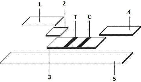
[0046] A test strip for quantitative detection of 25-hydroxyvitamin D, comprising a sample loading area 1, a conjugate release area 2, a reaction area 3 and a water absorption area 4 arranged in sequence; the conjugate release area is coated with mouse anti-human 25-hydroxyl A glass cellulose membrane of vitamin D monoclonal antibody, the mouse anti-human 25-hydroxyvitamin D monoclonal antibody is coated by colloidal gold, quantum dots or up-conversion luminescent material UCP; the reaction area is divided into a test area T and a control area C, The test area is coated with 25-hydroxyvitamin D-coupled protein, and the control area is a nitrocellulose membrane coated with goat anti-mouse IgG antibody.
[0047] Further, the quantitative detection test strip also includes a PVC backing plate (5), and the sample loading area, conjugate release area, reaction area, and water absorption area are sequentially overlapped and pasted on the PVC backing plate.
[0048] Further, the prep...
Embodiment 2
[0059] Example 2 : Preparation of the conjugate-releasing region
[0060] Add 2μmol colloidal gold (or quantum dots or up-conversion luminescent material UCP) and 1mg mouse anti-human 25-hydroxyvitamin D monoclonal antibody to 1mL pH7.4, 50mM phosphate buffer, shake gently at room temperature for 2 hours, then Centrifuge at 12000r / min for 30min, and take the precipitate.
[0061] Colloidal gold (or quantum dots or up-conversion luminescent material UCP) labeled mouse anti-human 25-hydroxyvitamin D monoclonal antibody, spread 20cm according to 1ml solution 2 Spray on the glass cellulose film with a film sprayer, place it at a temperature of 20-30°C, and dry it at a relative humidity of 30-35% for 5 hours to obtain a conjugate release area.
Embodiment 3
[0062] Example 3: Preparation of the reaction zone
[0063] Use 0.01M pH 7.4 phosphate buffer to prepare 25-hydroxyvitamin D-coupling protein into a solution of 0.8~1.6mg / ml, and coat it on a nitrocellulose membrane with a spray film instrument at a volume of 1~1.5μl / cm The area to be tested; another goat anti-mouse IgG antibody was formulated into a solution of 1-3 mg / ml, and the control area was coated with a film sprayer at an amount of 1-1.5 μl / cm; Dry at 38°C for 8-12 hours to prepare a nitrocellulose membrane coated with a detection area of 25-hydroxyvitamin D-coupled protein and a control area coated with goat anti-mouse IgG.
PUM
| Property | Measurement | Unit |
|---|---|---|
| diameter | aaaaa | aaaaa |
| particle diameter | aaaaa | aaaaa |
| wavelength | aaaaa | aaaaa |
Abstract
Description
Claims
Application Information
 - R&D
- Intellectual Property
- Life Sciences
- Materials
- Tech Scout
- Unparalleled Data Quality
- Higher Quality Content
- 60% Fewer Hallucinations
Browse by: Latest US Patents, China's latest patents, Technical Efficacy Thesaurus, Application Domain, Technology Topic, Popular Technical Reports.
© 2025 PatSnap. All rights reserved.Legal|Privacy policy|Modern Slavery Act Transparency Statement|Sitemap|About US| Contact US: help@patsnap.com


