Method for marking protein by carboxyl microsphere

A technology for labeling proteins and microspheres, which is applied in the direction of material inspection products, measuring devices, instruments, etc., can solve problems such as harsh reaction conditions, wasting antibodies, and affecting coupling efficiency, achieving high-efficiency coupling, reducing dosage, and avoiding hydrolysis Effect

Inactive Publication Date: 2015-09-09
BIOSINO BIO TECH & SCI
View PDF5 Cites 17 Cited by
  • Summary
  • Abstract
  • Description
  • Claims
  • Application Information

AI Technical Summary

Problems solved by technology

[0004] The advantage of the one-step method is that it is easy to operate. Although it is relatively simple to use EDC as a cross-linking agent, this method is uncontrollable. In addition to the target product, the final product may also have a large number of self-ligated products of multiple antibodies, which not only wastes antibodies. , also greatly reduces the reaction rate
The two-step method uses EDC and NHS or Sulfo-NHS as a cross-linking agent, because the reaction is carried out step by step, the first step is to activate the carboxyl group, and then add the antibody after washing the microspheres, so the direction of the reaction is certain and controllable There are two most commonly used methods for washing microspheres after activation. One is centrifugation. Even precipitation seriously affects the efficiency of coupling; the second is tangential flow filtration, which will not cause latex precipitation, but it takes a long time and affects the efficiency of the second step reaction; if it is magnetic carboxyl microspheres, it is relatively Convenience, beads can be washed using a magnetic separation system
The common disadvantage of the two chemical coupling methods is that the requirements for the reaction conditions are relatively harsh. For example, the one-step method must be reacted in an acidic environment, the first step of the two-step method must be in an acidic environment, and the second step is preferably in an acidic environment. In an alkaline environment

Method used


Image

  • Method for marking protein by carboxyl microsphere
  • Method for marking protein by carboxyl microsphere
  • Method for marking protein by carboxyl microsphere

Examples


Embodiment 1

[0050] Example 1 Carboxyl latex microspheres labeled cystatin C antibody

[0051] Cystatin C antibody is a product of Oriental Yeast Company, polyclonal antibody, the concentration is 15mg / mL. The antibody only reacts with human cystatin C and has no cross-immune reaction with other antigens, which basically meets the requirements of this experiment. The latex microspheres are products of JSR Company, the particle size is 61 nm, the carboxyl content is 0.190 mmol / g, the concentration is 5.2%, and the solution is purified water with 0.09% sodium azide. EDC and NHS are products of Pierce Company.

[0052] Latex microspheres are prepared into 0.5% solution with 10mM MES, the activation solution of pH 6.00, stir evenly, according to the carboxyl group content on the latex particle surface, according to the ratio of molar ratio EDC:NHS:-COOH=1:1:1, calculate all The required amount of EDC and NHS was weighed and directly added to the latex microsphere solution, stirred while addi...

Embodiment 2

[0058] The preparation and application of the assay kit of embodiment 2 cystatin C

[0059] (1) Prepare R2 reagent.

[0060] The latex particles (i.e. the carboxyl microspheres labeled cystatin C antibody prepared in Example 1) connected to the cystatin C antibody were washed with 10mM Tris, 0.1%BSA, 0.1%NaN 3 , Dilute the pH7.50 solution to 0.05% to make R2 reagent.

[0061] (2) Prepare R1 reagent.

[0062] Add 2.5% NaCl and 0.5% Tween-20 to a buffer solution with a final concentration of 2.5% and 0.5% Tween-20 in 10 mM Tris buffer solution with a pH value of 8.5, and stir evenly to obtain reagent R1.

[0063] (3) Preparation of standard products

[0064] Dissolve pure cystatin C in 10 mM Tris, 0.1% BSA, 0.1% NaN 3 , in a pH 7.50 solution, make standard products with gradient concentrations of about 0, 0.5, 1, 2, 4, and 8 mg / L.

[0065] Reagents R1, R2 and standards constitute a cystatin C assay kit.

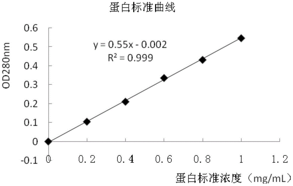
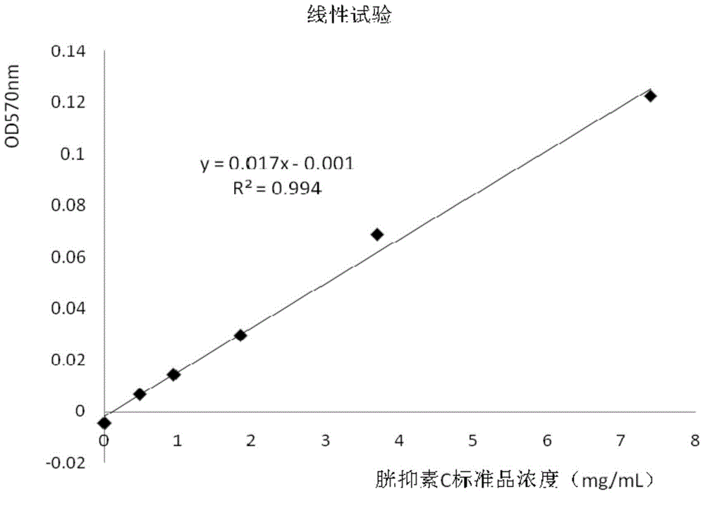
[0066] (4) Linearity test of cystatin C assay kit detection

[0067...

Embodiment 3

[0073] Example 3 Carboxyl Magnetic Microspheres Coupling Streptavidin

[0074] The magnetic microspheres are products of Merck Company, the particle size is 0.5-2 μm, the carboxyl content is 78 μmol / g, the concentration is 10%, and the solution is purified water with 0.09% sodium azide; Dry powder; EDC and NHS are products of Pierce Company.

[0075] The magnetic microspheres are prepared into a 1% solution with 20mM MES, pH 5.80 activation solution, stirred evenly, according to the carboxyl content on the surface of the magnetic microspheres, according to the ratio of molar ratio EDC:NHS:-COOH=1:1:1, calculate The required amount of EDC and NHS was weighed and directly added to the magnetic microsphere solution, stirred while adding, and stirred at room temperature for 15 minutes. Immediately use NaOH to adjust the pH value of the magnetic microsphere solution to 8.5, then add streptavidin dissolved in purified water in advance to make the final concentration of streptavidin...


PUM

PropertyMeasurementUnit
diameteraaaaaaaaaa

Abstract

The invention provides a method for marking protein by a carboxyl microsphere, belonging to the field of preparation of immunologic diagnosis kits. According to invention, in a system for marking the protein by the carboxyl microsphere, according to the content of carboxyls on the surface of the microsphere, the molar ratio of cross-linking agents EDC to -COOH is set to be (0.8-2) to 1, and the molar ratio of EDC to NHS or Sulfo to NHS is (1-3) to 1, so that the optimal ratio of the number of the carboxyls on the surface of the microsphere to the number of activated carboxyls is achieved; meanwhile, the pH is regulated, so that the reaction system is rapidly converted from condition beneficial for activation into state beneficial for antibody connection, the step of centrifugalization or tangential flow filtering is saved, and finally, the purposes of simplifying marking steps, saving cost, and marking protein by the carboxyl microsphere efficiently are achieved. The method provided by the invention can be applied to preparation of a detection kit taking the latex microsphere or the magnetic microsphere as a carrier, and has good market application prospect and economic value.

Description

technical field [0001] The invention relates to the technical field of preparation of immunodiagnostic kits, in particular to a novel, low-cost and easy-to-operate carboxyl microsphere-labeled protein method. Background technique [0002] Nano-microspheres are widely used in in vitro diagnostic reagents, which greatly improve the sensitivity of the reagents and can detect substances with minimal sample content. Nano-microspheres are used as carriers in diagnostic reagents with uniform size. The surface of the microspheres is coated with protein molecules such as antibodies and antigens. There are two methods for protein molecules to adsorb onto nano-microspheres: physical adsorption and chemical coupling. Physical adsorption is a relatively traditional and effective method, but it also has the disadvantages of being sensitive to system changes and poor stability; chemical coupling can avoid these defects. In a narrow sense, chemical coupling refers to the use of commerciall...

Claims


Application Information

Patent Timeline
Patent Type & Authority Applications(China)
IPC IPC(8): G01N33/68
CPCG01N33/6803
Inventor 黎霞赵文姬张志栋
Owner BIOSINO BIO TECH & SCI
Why Eureka
Try Eureka