
Solid tumor patient autologous NK cell separation, excitation, amplification and activity detection method

A technology for NK cells and solid tumors, applied in biochemical equipment and methods, animal cells, vertebrate cells, etc., to achieve the effects of multiple amplification, simple operation steps, and low cost of reagents and consumables

Active Publication Date: 2015-09-23
山西大医院
View PDF3 Cites 21 Cited by
  • Summary
  • Abstract
  • Description
  • Claims
  • Application Information

AI Technical Summary

Problems solved by technology

[0011] The present invention aims to overcome the deficiencies of the existing methods for the in vitro separation and culture, activation and expansion, and detection of cytotoxic activity of autologous NK cells from patients with solid tumors, and improve and optimize the steps of NK cell collection and separation, activation and expansion, and detection of cytotoxic activity.

Method used


Image

  • Solid tumor patient autologous NK cell separation, excitation, amplification and activity detection method
  • Solid tumor patient autologous NK cell separation, excitation, amplification and activity detection method
  • Solid tumor patient autologous NK cell separation, excitation, amplification and activity detection method

Examples


Embodiment 1

[0071] A method for the isolation, activation and expansion of autologous NK cells from patients with solid tumors, comprising the following steps:

[0072] (1) Collection and isolation of NK cells:

[0073] 1.1. Peripheral blood collection: 40-60ml of peripheral blood from patients with solid tumors, preferably 50ml. The collection device is a disposable 100ml syringe, which is filled with 10ml of normal saline and 50unit / ml of heparin sodium anticoagulant, and the dosage of heparin sodium is calculated according to the volume of blood collected. Take 2ml of peripheral blood and detect lymphocyte subsets by flow cytometry.

[0074] 1.2. NK cell separation:

[0075] 1.2a. Put the collected peripheral blood into a 50ml centrifuge tube, centrifugation conditions: centrifugal force 500g, centrifugation time 10min. Aspirate the plasma with a straw and put it into another centrifuge tube, inactivate it in a water bath at 56°C for 30 minutes and then centrifuge. Centrifugation co...

Embodiment 2

[0106] A method for detecting cytotoxicity of autologous NK cells of patients with solid tumors, comprising the steps of:

[0107] 3.1. CFSE labeling target cells:

[0108] Tumor cell culture: suspend the tumor cell line (K562 cells) in 1640 medium containing 10% inactivated fetal bovine serum (FBS), and place at 37°C, 5% CO 2 , subcultured under saturated humidity conditions.

[0109] CFSE-labeled target cells: collect tumor cells in good growth condition, use PBS buffer, centrifuge at 300g centrifugal force, and centrifuge at room temperature for 5min. Wash twice, count with trypan blue and resuspend cells with PBS buffer. Add the pre-warmed CFSE-PBS solution into the cell mass (the final concentration of CFSE is 2umol / L), gently blow and beat the suspended cells (cell concentration 1×10 6 / mL). Place the cells at 37°C for 5-10min. Add 10 mL of 1640 medium containing 10% FBS to terminate the reaction. Centrifuge the cells, add 10 mL of 1640 medium containing 10% FBS, pla...


PUM


Abstract

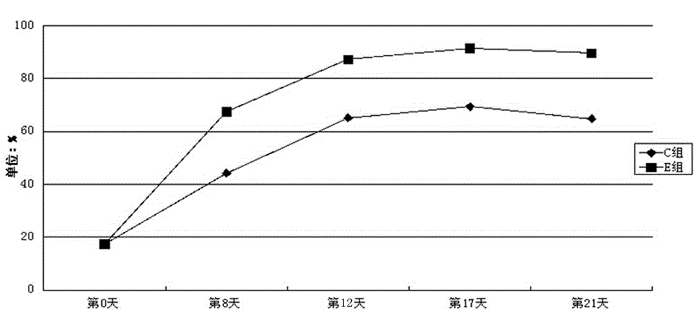
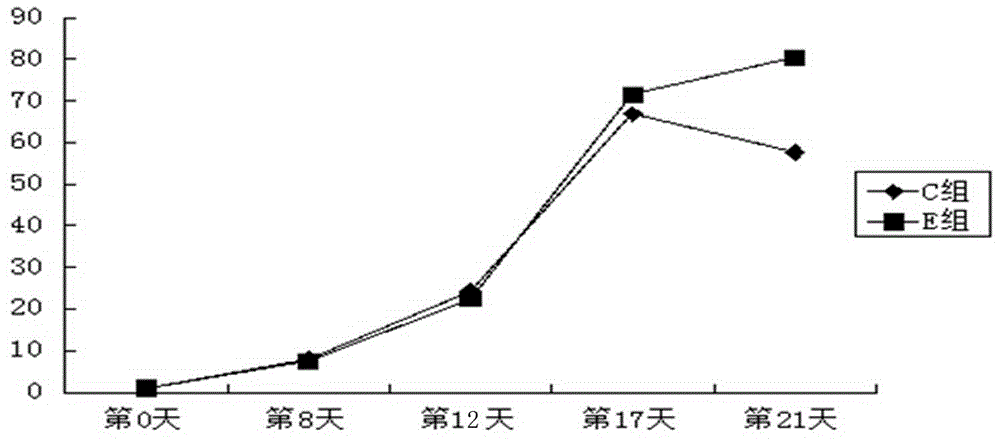
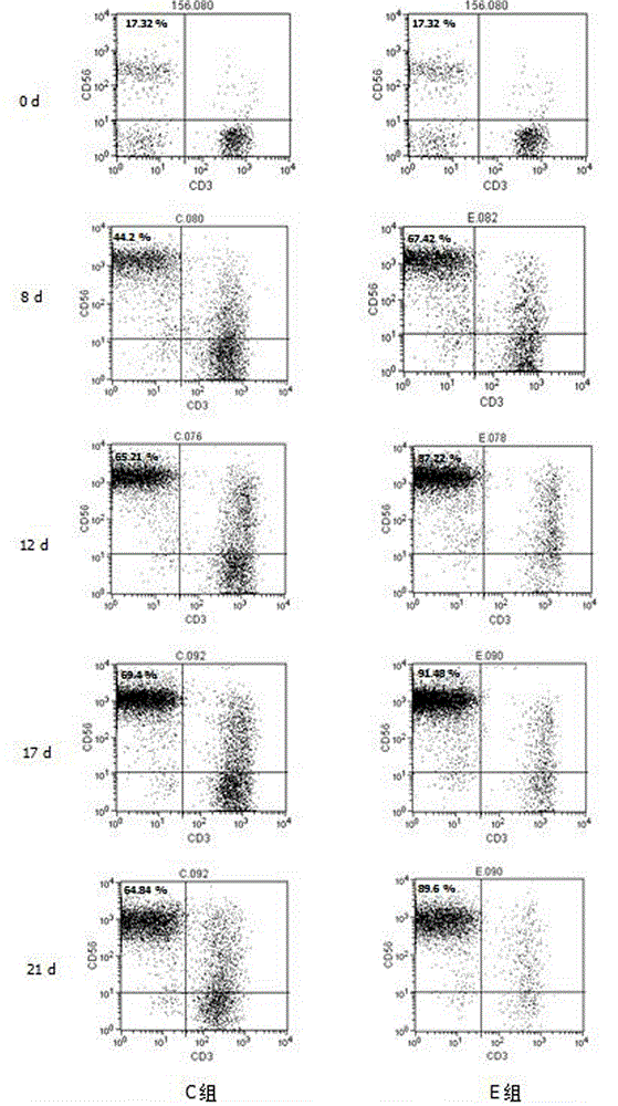
The invention provides a solid tumor patient autologous NK cell separation, excitation, amplification and activity detection method. By the adoption of the NK cell separation, culturing and amplification method, highly active lymphocyte populations can be obtained through separation from peripheral blood, through stimulation of a series of in-vitro cell factors, NK cells (CD3-CD56+) are purified, activated and amplified greatly, and the NK cells can be applied to clinic treatment of tumors. Through the NK cell activity detection method, the kill rate of effector cells to target cells can be specifically analyzed in mixed cell sap of immune cells and tumor cells, interference from the effector cells in a detection result is avoided, and early apoptosis target cells and late apoptosis target cells can be analyzed. The method is easy and convenient to implement, the flexibility is high, the kill function of a cell can be detected at the single cell level, and the method has important significance in fundamental research conducted before clinical application of tumor adoptive cellular immunotherapy and quality control conducted in the clinical application process.

Description

technical field [0001] The technology of the present invention mainly relates to a method for in vitro separation and culture, activation and amplification, and detection of cytotoxic activity of NK cells derived from autologous peripheral blood of patients with solid tumors. Background technique [0002] Natural killer cells (NK), also known as natural killer cells, have anti-virus infection and anti-tumor without MHC restriction, and have been used as an important therapeutic method for clinical cell immunotherapy of tumors. As the body's natural immune cells, NK cells mainly kill tumor cells, viruses and clear non-self cells by directly killing tumor cells and secreting cytokines to regulate other immune cells. Rosenberg et al. found in the process of treating tumors with IL-2-activated LAK cells that the cells characterized by CD3-CD16+ cells (natural killer cells, NK) play the main role in anti-tumor, and NK cells are the main effector cells. Since then, researchers ha...

Claims


Application Information

Patent Timeline
IPC IPC(8): C12N5/0783C12Q1/02
Inventor 张俊萍郭继强韩亚萍贾原赵晴李芳
Owner 山西大医院
Learn More