Biological electron microscope sample carbonization method

A bioelectricity and sample technology, applied in the preparation of test samples, etc., can solve the problems of inability to observe, incapable of sampling, incapable of drying, etc., and achieve the effect of small chemical and physical shrinkage, no change in morphology and structure, and clear microstructure

Active Publication Date: 2015-11-04
FUDAN UNIV
View PDF2 Cites 3 Cited by
  • Summary
  • Abstract
  • Description
  • Claims
  • Application Information

AI Technical Summary

Problems solved by technology

However, some samples are restricted by the critical point conditions, and the critical point preparation conditions of the samples cannot be carried out, and the scanning electron microscope observation cannot be performed.
Such as: mixture of hydrogenated castor oil and fatty alcohol polyoxyethylene ether (21), mixture

Method used


Image

  • Biological electron microscope sample carbonization method
  • Biological electron microscope sample carbonization method
  • Biological electron microscope sample carbonization method

Examples


Embodiment 1

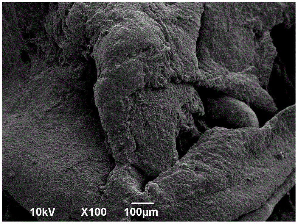
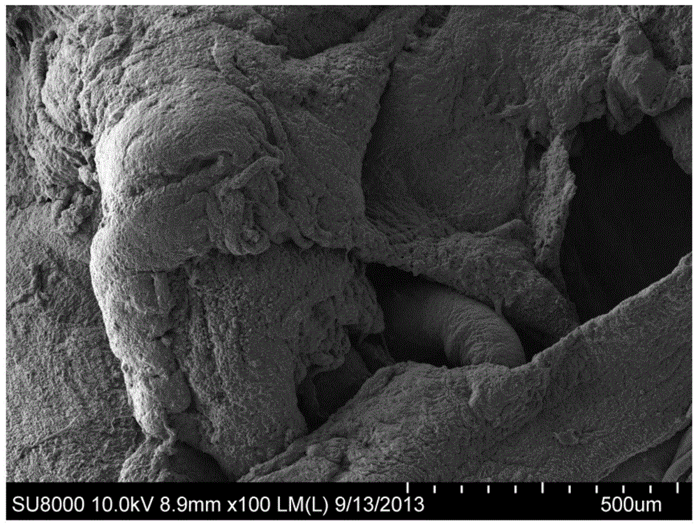
[0060] Example 1 Preparation of biological scanning electron microscope samples:

[0061] 1) Take isolated animal or human organ tissue blocks, after routine treatment, quickly fix with 2.5% glutaraldehyde 2ml for 2 hours or longer, refrigerate and fix at 4oC for more than 2 hours, rinse with buffer twice, 10 minutes each time ;

[0062] 2) Take the electron microscope nickel mesh as a support, remove the surface water stains of the tissue block, and place it on the electron microscope nickel mesh;

[0063] 3) Take a centrifuge tube with 1% osmium tetroxide solution added to the bottom, and place it horizontally to form a liquid swelling force at the bottom;

[0064] 4) Transfer the organized nickel mesh bracket to the middle or mouth end of the centrifuge tube, seal the centrifuge tube and seal with a parafilm, mark the sample nature and sample number, and put multiple centrifuge tube samples into a unified container , seal again, and mark the carbonization start time;

[...

Embodiment 2

[0069] Example 2 Preparation of Cell Scanning Electron Microscope Samples

[0070] 1) Take the separated red blood cells in a 1.5ml EP centrifuge tube, add 2.5% glutaraldehyde for discrete mixing, place in a 4oC refrigerator for 1-2 hours, centrifuge at 500-800 rpm, extract the supernatant fixative, add buffer Dispersed, mixed and rinsed, then centrifuged at 500-800 revolutions per minute, and the supernatant fixed solution was extracted, and repeated 2 times. Send samples in the state of suspension;

[0071] 2) Use a pipette to take an appropriate amount of suspended red blood cell solution, drop it on the grid containing the carbon film to settle for 3-5 minutes, and use filter paper to absorb the excess residual liquid on the grid to maintain humidity;

[0072] 3) Take a 1ml or 1ml EP centrifuge tube, add 100ul of 1% osmium tetroxide solution to the bottom of the centrifuge tube, the swelling force of the solution will make the solution deposit at the bottom of the EP cent...

Embodiment 3

[0080] Example 3 Preparation of Bacteria Scanning Electron Microscope Samples

[0081] 1) Put the accumulated bacteria into a 1.5ml EP centrifuge tube, select a mixed fixative of paraformaldehyde and glutaraldehyde, suspend and fix the accumulated bacteria at 4oC for more than 2 hours, centrifuge to remove the supernatant fixative, and use phosphate buffer The solution was mixed and centrifuged for rinsing, rinsing twice for 10 minutes each time, after removing the supernatant, add deionized distilled water and mix evenly to make the bacteria in a suspended state;

[0082] 2) Take a 1ml EP centrifuge tube and add 50ul-100ul of 0.1% osmium tetroxide to the 1ml EP centrifuge tube to form a liquid swelling force and keep it in the 1ml EP centrifuge tube;

[0083] 3) Take the carbon-containing membrane carrier net, pinch the edge of the carrier net with tweezers to suspend the carrier net, drop a small amount of bacterial suspension on one side of the carbon-containing film, stay ...


PUM


Abstract

The invention belongs to the field of biological sample preparation, and particularly relates to a biological electron microscope sample carbonization method. Volatile gas of 0.1 M osmium tetroxide liquid is adopted for conducting fumigation and burning on organic biological samples, so that the organic biological samples are completely carbonized to meet the requirement for scanning electron microscope observation. Biological electron microscope samples manufactured through the method have a better expression result and are environmentally friendly and low in carbon, operation is easy and convenient, the chemical and physical contraction rate of all the samples is small, the appearance structure is almost unchanged, and the fine structure is clear. The method breaks through the bottleneck of manufacturing of some electron microscope samples so that some transmission electron microscope samples and scanning electron microscope samples which can not be manufactured in the prior art can be smoothly manufactured and meet the requirement for electron microscope observation. The method can be used for biological electron microscope sample manufacturing of biological sample tissue, bacteria, organic macromolecule protein bodies or organic macromolecule substances.

Description

technical field [0001] The invention belongs to the field of biological sample preparation, in particular to a carbonization method for biological electron microscope samples Background technique [0002] Since the birth of the electron microscope, researchers have continuously explored and improved the pre-preparation, sectioning, and staining techniques of biological samples used for electron microscope observation in order to obtain higher-quality real images and experimental results. [0003] Conventional transmission bioelectron microscopy samples are prepared by embedding section method, negative staining method and so on. However, conventional scanning electron microscope sample preparation often adopts a single critical point drying method (critical point instrument freeze-drying method, also known as freeze-drying method). 2ml, fix for 2 hours or longer, rinse 3 times with 0.1M phosphate buffer, 15 minutes each time, then fix with 2ml of 1% osmium tetroxide for 2 h...

Claims


Application Information

Patent Timeline
IPC IPC(8): G01N1/28
Inventor 高鸿建
Owner FUDAN UNIV
Why Eureka
Try Eureka