Test paper for detecting metalaxyl residue and application thereof

A metalaxyl and test paper technology, which is applied in the field of test paper for the detection of metalaxyl residues, can solve the problems of tedious and time-consuming test sample processing, and achieve the effects of short detection time, wide application range and simple operation

Inactive Publication Date: 2016-01-20
贵州勤邦食品安全科学技术有限公司
1 Cites 19 Cited by

AI-Extracted Technical Summary

Problems solved by technology

[0004] From the perspective of existing technologies and related testing standards, the current detection methods for metalaxyl residues are mainly instrumental methods, and the most commonly used methods are LC-UV, LC-MS and LC-MS/MS. The instrumental methods have high detection sensitivity, Strong specificity and other advantages, but the pretreatment of the test sample is cumbersome and time-consuming, and the sample needs to be extracted and purified. At the ...

Abstract

The invention discloses a test paper for detecting metalaxyl residue and application thereof. The test paper comprises a sample absorption pad (1), a conjugate release pad (2), a reaction membrane (3), a water absorption pad (4) and a base plate (7), wherein the reaction membrane comprises a detection line (5) coated with a metalaxyl semiantigen-carrier protein conjugate and a quality control line (6) coated with a goat anti-mouse antiantibody, and the conjugate release pad (2) is coated with a metalaxyl monoclonal antibody-colloidal gold marker. The invention also provides a method for applying the metalaxyl test paper in detection of metalaxyl residue in tea, tobacco and vegetable. The test paper provided by the invention can be used for detecting metalaxyl residue in tea, tobacco and vegetable, has the characteristics of simple operation, high sensitivity, a fast detection speed, low cost, etc., and is applicable to screening and on-site monitoring of a great number of samples.

Application Domain

Technology Topic

MetalaxylChemistry +6

Image

  • Test paper for detecting metalaxyl residue and application thereof
  • Test paper for detecting metalaxyl residue and application thereof
  • Test paper for detecting metalaxyl residue and application thereof

Examples

  • Experimental program(3)

Example Embodiment

[0040] The preparation of embodiment 1 metalaxyl detection test paper
[0041] The preparation method of the test paper mainly includes the following steps:
[0042] 1) Prepare a conjugate release pad coated with a metalaxyl monoclonal antibody-colloidal gold marker;
[0043] 2) Prepare a reaction membrane with a detection line coated with a metalaxyl hapten-carrier protein conjugate and a quality control line coated with a goat anti-mouse antibody;
[0044] 3) Assemble the conjugate release pad, reaction membrane, sample absorption pad, water absorption pad and PVC bottom plate prepared in 1) and 2) into a test paper.
[0045] The following is a step-by-step detailed description:
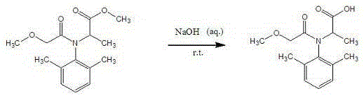
[0046] 1. Preparation of metalaxyl hapten
[0047] Metalaxyl was dissolved in ethanol, 10% NaOH aqueous solution was added with stirring at room temperature, and the reaction was monitored by TLC until there was no raw material or the raw material point was very shallow. The reaction was stopped, purified, and recrystallized from methanol to obtain the hydrolyzate. , infrared and mass spectrometry identification, metalaxyl hapten was successfully synthesized.
[0048] 2. Immunogen preparation - Metalaxyl hapten is coupled with bovine serum albumin (BSA) to obtain immunogen.
[0049] Weigh 26.5mg hapten and dissolve it in 2mL DMF solution, add 60mg EDC and NHS (dissolved in 2mL water) for activation for 30 minutes, add to 110-264mg carrier protein BSA (dissolve in 5mL water) for coupling to prepare immunogen, Dialyze with 0.02mol/LPB buffer for 3 days, and change the dialysate every morning and evening to remove unreacted small molecular substances; after the dialysis is completed, it is used for animal immunization to prepare antibodies. Aliquot and store at -20°C for later use.
[0050] 3. Preparation of the original coating
[0051] Dissolve 12 mg of metalaxyl hapten in 0.8 ml of DMF, and cool it to 10 °C to obtain reaction solution I; add 10 ul of isobutyl chloroformate to I, and stir at 10 °C for 30 min; take 30 mg of bovine serum albumin with 2.2 ml of 50 mmol/LNa 2 CO 3 Dissolve, react at 10°C for 4h, and then overnight at 4°C; dialyze with 0.01mol/L PBS at 4°C for 3 days, and change the dialysate 3 times a day to remove unreacted small molecular substances to obtain the metalaxyl coating, which is divided into Store at -20°C for later use.
[0052] 4. Preparation method of metalaxyl monoclonal antibody
[0053] (1) Animal immunity
[0054] The immunogen obtained in step 2 was injected into Balb/c mice at an immunizing dose of 150 μg/mouse to produce antiserum.
[0055] (2) Cell fusion and cloning
[0056] After the mouse serum test results were higher, the spleen cells were taken and fused with SP2/0 myeloma cells in a ratio of 8:1 (quantity ratio), and the cell supernatant was determined by indirect competitive ELISA, and the positive wells were screened. The positive wells were cloned by limiting dilution method until a hybridoma cell line secreting metalaxyl monoclonal antibody was obtained.
[0057] Cell cryopreservation and recovery: The monoclonal hybridoma cell line was made into a cell suspension of 1×10⁶ cells/ml with a cryopreservation solution, and stored in liquid nitrogen for a long time. When resuscitated, take out the cryopreservation tube, immediately put it into a 37°C water bath to thaw quickly, remove the cryopreservation solution by centrifugation, and transfer it to a culture bottle for culture.
[0058] Production and purification of monoclonal antibodies: Balb/c mice were intraperitoneally injected with sterilized paraffin oil 0.5ml/mouse, and 5×10⁵/mouse of stable monoclonal hybridoma cell lines were intraperitoneally injected 7 days later, and ascites was collected 7 days later. The ascites was purified by the octanoic acid-saturated ammonium sulfate method and stored at -20°C.
[0059] (3) Cell cryopreservation and recovery
[0060] Hybridoma cells were made into a cell suspension of 1×10⁶ cells/ml with a cryopreservation solution, and stored in liquid nitrogen for a long time. When resuscitated, take out the cryopreservation tube, immediately put it into a 37°C water bath to thaw quickly, remove the cryopreservation solution by centrifugation, and transfer it to a culture bottle for culture.
[0061] (4) Preparation and purification of monoclonal antibodies
[0062] Incremental culture method: The hybridoma cells were placed in the cell culture medium, cultured at 37°C, and the obtained culture solution was purified by the octanoic acid-saturated ammonium sulfate method to obtain monoclonal antibodies, which were stored at -20°C.
[0063] The cell culture medium is to add calf serum and sodium bicarbonate to the RPMI1640 medium, so that the final concentration of calf serum in the cell culture medium is 20% (mass percentage), so that the sodium bicarbonate is in the cell culture medium. The final concentration in 0.2% (mass percentage); the pH of the cell culture medium was 7.4.
[0064] 5. Preparation of goat anti-mouse anti-antibodies
[0065] The sheep were used as immunized animals, and the mouse-derived antibodies were used as immunogens to immunize pathogen-free sheep to obtain goat anti-mouse anti-antibodies.
[0066] 6. Preparation of monoclonal antibody-colloidal gold label
[0067] (1) Preparation of colloidal gold
[0068] Dilute 1% chloroauric acid to 0.01% (mass percentage) with double-distilled deionized water, take 100ml and place it in a conical flask, heat it to boiling with a constant temperature electromagnetic stirrer, and add 2.5% under continuous high temperature and continuous stirring. ml 1% trisodium citrate, continue to stir and heat at a constant speed until the solution turns bright red, stop, cool to room temperature, restore to the original volume with deionized water, and store at 4°C. The prepared colloidal gold appears pure, translucent, free of precipitation and floating matter.
[0069] (2) Preparation of gold-labeled antibodies
[0070] Under magnetic stirring, adjust the pH of colloidal gold to 7.0 with 0.2 mol/L potassium carbonate solution, add metalaxyl antibody to the colloidal gold solution according to the standard of adding 20-50 μg antibody per ml of colloidal gold solution, and continue to stir and mix. Homogenize for 30 min, add 10% BSA to make the final concentration in the colloidal gold solution 1% (volume percentage), and let stand for 10 min. Centrifuge at 12,000 r/min and 4°C for 40 min, discard the supernatant, wash the precipitate twice with reconstitution buffer, and resuspend the precipitate with reconstitution buffer whose volume is 1/10 of the initial volume of colloidal gold, and store at 4°C for later use.
[0071] Reconstitution buffer: 0.02mol/L phosphate containing bovine serum albumin (BSA) 0.1%-0.3% (volume percentage), Tween-800.05%-0.2% (mass percentage), pH 7.2 buffer.
[0072] 7. Preparation of conjugate release pads
[0073] Soak the conjugate release pad in phosphate buffer containing bovine serum albumin (the concentration of bovine serum albumin in the buffer is 0.5%) and pH 7.2, 0.5mol/L phosphate buffer, soak it uniformly for 1h, and bake it at 37℃ 3h spare. Using the ZX1000 film spraying instrument produced by Baidao Company, the prepared metalaxyl monoclonal antibody-colloidal gold label was uniformly coated on the conjugate release pad, and each 1 cm of the conjugate release pad was coated with 0.01ml of metalaxyl monoclonal antibody. - After the colloidal gold marker, put it in a 37°C environment (humidity <20%) for 60 minutes, take it out, seal it and store it in a dry environment (humidity <20%) for later use.
[0074] 8. Preparation of reaction membrane
[0075] The metalaxyl hapten-bovine serum albumin conjugate was coated on the reaction membrane to form the detection line (T line), and the goat anti-mouse antibody was coated on the reaction membrane to form the quality control line (C line).
[0076] Coating process: Dilute the metalaxyl hapten-bovine serum albumin conjugate to 10 mg/ml with phosphate buffer, and coat it on the detection line on the nitrocellulose membrane with the ZX1000 type membrane instrument produced by Biodoor (T line), the coating amount is 1.0μg/cm 2; Dilute the rabbit anti-goat anti-antibody to 200 μg/ml with 0.01mol/L, pH 7.4 phosphate buffer, and coat it on the quality control line (C line) on the nitrocellulose membrane with a dot membrane instrument, The coating amount is 1.0μg/cm 2. The coated reaction membrane was dried at 37 °C for 2 h and used for later use.
[0077] 9. Preparation of sample absorbent pads
[0078] The sample absorption pad was soaked in phosphate buffer containing 0.5% bovine serum albumin (volume percentage) and pH 7.20.1 mol/L for 2 hours, and then dried at 37 °C for 2 hours.
[0079] 10. Assembly of test strips
[0080] Paste the sample absorption pad, the conjugate release pad, the reaction film, and the water absorbent pad on the PVC bottom plate in sequence; 1/3 of the area of ​​the conjugate release pad is covered by the sample absorption pad from the starting end, and the end of the conjugate release pad is connected with the reaction pad. The beginning of the membrane is connected, the end of the reaction membrane is connected to the beginning of the absorbent pad, the beginning of the sample absorbing pad is aligned with the beginning of the PVC bottom plate, and the end of the absorbent pad is aligned with the end of the PVC bottom plate; there are detection lines and quality control on the reaction membrane. Line, detection line (T line) and quality control line (C line) are all strips perpendicular to the length of the test paper; the detection line is located on the side near the end of the conjugate release pad; the quality control line is located away from the binding One side of the end of the release pad; cut the test paper into 3mm wide strips by machine, and put it in a special plastic card to form a test paper card, which can be stored for 12 months at 4-30 °C.

Example Embodiment

[0081] The detection of metalaxyl residue in the sample of embodiment 2
[0082] 1. Sample pretreatment
[0083] Take 1.0g±0.05g of homogeneous sample into a 50ml polystyrene centrifuge tube, add 5ml of extract, shake with a shaker for 5min, centrifuge at 3000g for 5min at room temperature, and take the supernatant for testing.
[0084] 2. Test with test paper
[0085] Use a pipette to suck up the sample solution to be tested and add 2-3 drops vertically to the sample addition hole. When the liquid flows, start timing and react for 5 minutes. The result is judged, and other times are judged invalid.
[0086] 3. Analysis of test results
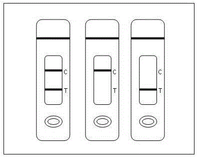
[0087] Negative (-): Both the T line and the C line are colored, indicating that the metalaxyl concentration in the sample is below the detection limit, such as image 3.
[0088] Positive (+): T line has no color and C line has color, indicating that the drug concentration of metalaxyl in the sample is equal to or higher than the detection limit, such as image 3.
[0089] Invalid: No C line appears, indicating incorrect operation process or the test strip has deteriorated and failed, such as image 3. In this case, the instructions should be read carefully again and the test should be re-tested with a new test strip.

Example Embodiment

[0090] Example 3 Sample Detection
[0091] 1. Detection limit test
[0092] A blank tobacco sample was taken, and metalaxyl was added to it to a final concentration of 1, 2, and 4 mg/kg, respectively. Test paper was taken for detection, and the measurement was repeated three times for each sample.
[0093] When testing tobacco, tea, and vegetable samples with test paper, when the metalaxyl concentration is 1mg/kg, the test paper shows two red lines visible to the naked eye, which is negative; when the metalaxyl concentration is 2, 4mg /kg, the quality control line of the test paper is displayed, but the detection line is not displayed, which is positive; it indicates that the detection line of this test paper to metalaxyl is 2 mg/kg.
[0094] 2. False positive rate and false negative rate test
[0095] Take 20 positive tobacco samples with known metalaxyl content greater than 2 mg/kg and 20 negative tobacco samples with known metalaxyl content less than 2 mg/kg, and use three batches of test paper to detect the negative and positive rates. The results are shown in Table 1 and Table 2.
[0096] Table 1 Test positive sample results
[0097]
[0098] Table 2 Test results of negative samples
[0099]
[0100]The results show that when testing positive samples with the test strips produced in 3 batches, all the results are positive. It can be seen that the coincidence rate of positive samples is 100%, and the false negative rate is 0. When testing 20 negative samples, there is one false positive in one batch. The test results showed that the negative coincidence rate was 98%, and the false positive rate was less than 5%. It is illustrated that the test paper for detecting metalaxyl residues of the present invention can rapidly detect metalaxyl residues in tobacco.
[0101] 3. Specificity test
[0102] Specificity is often expressed as a cross-reactivity ratio, which refers to the ability of an antibody to bind to antigenic determinants with different structures. The samples of 500 μg/L of carbendazim, triadimefon and pyrethroids that were routinely tested were detected with metalaxyl colloidal gold test paper. The results showed that when the test paper was used to detect 500 μg/L of carbendazim, triadimefon and pyrethroids, the quality control line and detection line of the test paper were both colored and negative, indicating that the test paper had no cross-reaction to these drugs.

PUM

no PUM

Description & Claims & Application Information

We can also present the details of the Description, Claims and Application information to help users get a comprehensive understanding of the technical details of the patent, such as background art, summary of invention, brief description of drawings, description of embodiments, and other original content. On the other hand, users can also determine the specific scope of protection of the technology through the list of claims; as well as understand the changes in the life cycle of the technology with the presentation of the patent timeline. Login to view more.

Similar technology patents

Integrated microfluidic optical device for sub-micro liter liquid sample microspectroscopy

InactiveUS20090051901A1Great sensitivityHigh sensitivityRadiation pyrometrySpectrum investigationSpectroscopySpectroscopic detection
Owner:DYNAMIC THROUGHPUT

Classification and recommendation of technical efficacy words

  • High sensitivity
  • Strong specificity

System and method for optical coherence imaging

ActiveUS20060055936A1Sufficient signal to noise ratioHigh sensitivityInterferometersScattering properties measurementsPhysicsCoherent imaging
Owner:THE GENERAL HOSPITAL CORP

Catheter-free implantable needle biosensor

ActiveUS20080033269A1High sensitivitySimple stepSurgeryVaccination/ovulation diagnosticsNeedle electrodeElectricity
Owner:VIRTUE MEDICAL TECH (HUZHOU) CO LTD

Cas9-foki fusion proteins and uses thereof

ActiveUS20150071899A1Strong specificityReduce the possibilityFusion with DNA-binding domainPeptide/protein ingredientsResearch settingNuclease
Owner:PRESIDENT & FELLOWS OF HARVARD COLLEGE

c-MET MODULATORS AND METHOD OF USE

InactiveUS20070179130A1Strong specificityEliminate side effectsBiocideSenses disorderSignal transductionCellular activity
Owner:EXELIXIS INC
Why Eureka
Try Eureka