Method for isolated culture of esophagus epithelial stem cells

A technology for separating and culturing stem cells, applied in the field of cell biology, which can solve the problems of unresearched methods of separating and culturing esophageal epithelial stem cells, and achieve the effects of prolonging cell activity, inhibiting multi-directional differentiation potential, and stabilizing heredity

Active Publication Date: 2016-04-20
THE FIRST AFFILIATED HOSPITAL OF HENAN UNIV OF SCI & TECH
View PDF2 Cites 4 Cited by
  • Summary
  • Abstract
  • Description
  • Claims
  • Application Information

AI Technical Summary

Problems solved by technology

[0004] Studies have shown that there are many studies on primary culture methods of human esophageal ep

Method used


Image

  • Method for isolated culture of esophagus epithelial stem cells
  • Method for isolated culture of esophagus epithelial stem cells
  • Method for isolated culture of esophagus epithelial stem cells

Examples


Embodiment 1

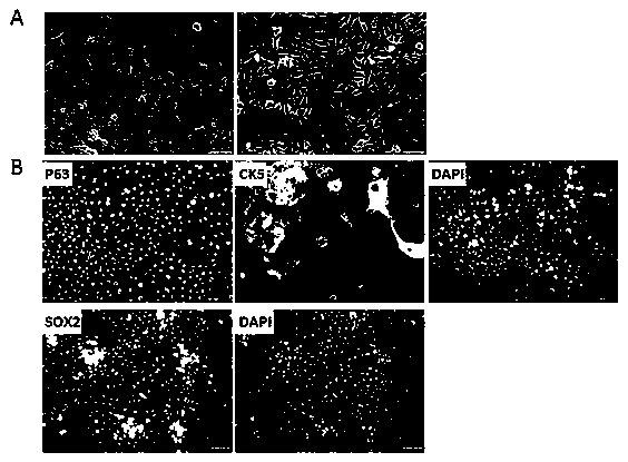
[0029] Isolation, culture and identification of human esophageal epithelial stem cells

[0030] 1.1 Isolation and culture

[0031] (1) Materials: Discarded normal epithelial tissue of the esophagus of a 69-year-old male was removed under aseptic conditions, and placed in phosphate-buffered saline (PBS) / normal saline solution containing penicillin 100ug / mL and streptomycin 100ug / mL In a sterile tube, take it back to the laboratory immediately;

[0032] (2) Separation: Wash the esophageal epithelial tissue 3 to 5 times with PBS containing 200 μg / mL penicillin / streptomycin, and remove hemorrhagic tissue, necrotic tissue and fibrovascular tissue; Scissors and ophthalmic forceps to trim the tissue to 0.5-1mm 3 Then add the centrifuge tube containing the digestion solution of 0.1% collagenase type I, 0.1% pronase and 0.5 mg / ml deoxyribonuclease I, and then put the centrifuge tube into a 37°C incubator for constant temperature digestion for 2h Afterwards, the digestion mixture was...

Embodiment 2

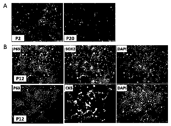
[0052] Isolation, culture and identification of mouse esophageal epithelial stem cells

[0053] 1. Cell isolation and culture

[0054] All animal studies were performed in full compliance with laboratory-grade animal standards. The normal esophageal tissues were taken out from the mice and washed three times with PBS containing antibiotics. Under sterile conditions, use sterile ophthalmic scissors and ophthalmic forceps to remove the hemorrhage and necrotic tissue in the esophagus, and cut them into pieces of 0.5-1mm 3 The small pieces were then added to the centrifuge tube containing the digestion solution of 0.1% pronase and 0.5 mg / ml DNase I. Digest at a constant temperature of 37°C for 3 hours, filter and centrifuge, and collect the cell pellet.

[0055] Resuspend the cells with the culture medium and place them in the culture bottle / dish covered with coating solution for horizontal culture, and culture the culture bottle / dish with EBSS containing 0.3mg / mL BSA and 40ug / ...


PUM


Abstract

The invention discloses a method for isolated culture of esophagus epithelial stem cells. The method comprises the steps of taking esophagus mucosa tissue and epithelium adhering to the esophagus mucosa tissue, removing muscular tissue, taking a basal layer of epithelium mucosae tissue, shearing the basal layer of epithelium mucosae tissue into bits, conducting digestion centrifugation, and spreading obtained cells in a culture flask, culture plate or culture dish coated with a matrix; then adding a DMEM/Ham's F-12 culture solution containing HEPES, sodium bicarbonate, L-glutamic acid, FBS, EGF, insulin, hydrocortisone, vibrio cholerae toxin, transferring, BPE, quasi-vitamin A and cell signaling pathway inhibitor; finally, placing the culture flask, culture plate or culture dish containing cell suspension in a 37 DEG C incubator for culture, and obtaining the single-layer epithelial stem cells in 10-14 days. According to the method, operation is convenient, safety performance is high, the esophagus epithelial stem cells are obtained through separation, the activity of the stem cells is high, and application prospects in tissue engineering and regenerative medicine are broad.

Description

technical field [0001] The invention belongs to the technical field of cell biology and relates to a stem cell isolation and culture technology, in particular to a method for the isolation and culture of esophageal epithelial stem cells. Background technique [0002] Stem cells are a type of pluripotent cells with self-replicating ability. Under certain conditions, it can differentiate into a variety of functional cells. Stem cells are divided into embryonic stem cells (EmbryonicStemCells, ESCs) and adult stem cells (AdultStemCells, ASCs) according to the developmental stage of stem cells. The proliferation and differentiation of embryonic stem cells constitute the basis of individual development, from a single fertilized egg to an individual with various tissues and organs; while adult stem cells can further differentiate into complex tissues and organs, and its research involves almost all life sciences and biological The field of medicine has bright prospects for develo...

Claims


Application Information

Patent Timeline
IPC IPC(8): C12N5/074
CPCC12N5/0602C12N2500/05C12N2500/25C12N2500/32C12N2500/38C12N2500/60C12N2500/84C12N2501/01C12N2501/11C12N2501/15C12N2501/155C12N2501/727C12N2501/998C12N2501/999C12N2509/00
Inventor 高社干冯笑山梁硕牟红梅刘刚原翔马志坤孔金玉
Owner THE FIRST AFFILIATED HOSPITAL OF HENAN UNIV OF SCI & TECH
Why Eureka
Try Eureka