Simple and green preparation method for monodisperse core-shell-structured C-dots@SiO2 fluorescence imaging nanoprobe

A technology of fluorescence imaging and core-shell structure, which is applied in the field of nanocomposite materials and its applications, can solve the problems of complex synthesis methods, cytotoxicity, and toxic drugs, and achieve simple synthesis methods, easy centrifugation and washing, and good biocompatibility sexual effect

Inactive Publication Date: 2016-06-01
NORTHEAST NORMAL UNIVERSITY
View PDF1 Cites 5 Cited by
  • Summary
  • Abstract
  • Description
  • Claims
  • Application Information

AI Technical Summary

Problems solved by technology

Nowadays, there are also a lot of SiO 2 The method of coating fluorescent materials to prepare fluorescent nanoprobes, but they have some disadvantages, such as complex synthesis methods, toxic drugs used in the synthesis process, pollution to the environment and certain toxic effects on cells

Method used


Image

  • Simple and green preparation method for monodisperse core-shell-structured C-dots@SiO2 fluorescence imaging nanoprobe
  • Simple and green preparation method for monodisperse core-shell-structured C-dots@SiO2 fluorescence imaging nanoprobe
  • Simple and green preparation method for monodisperse core-shell-structured C-dots@SiO2 fluorescence imaging nanoprobe

Examples


Embodiment 1

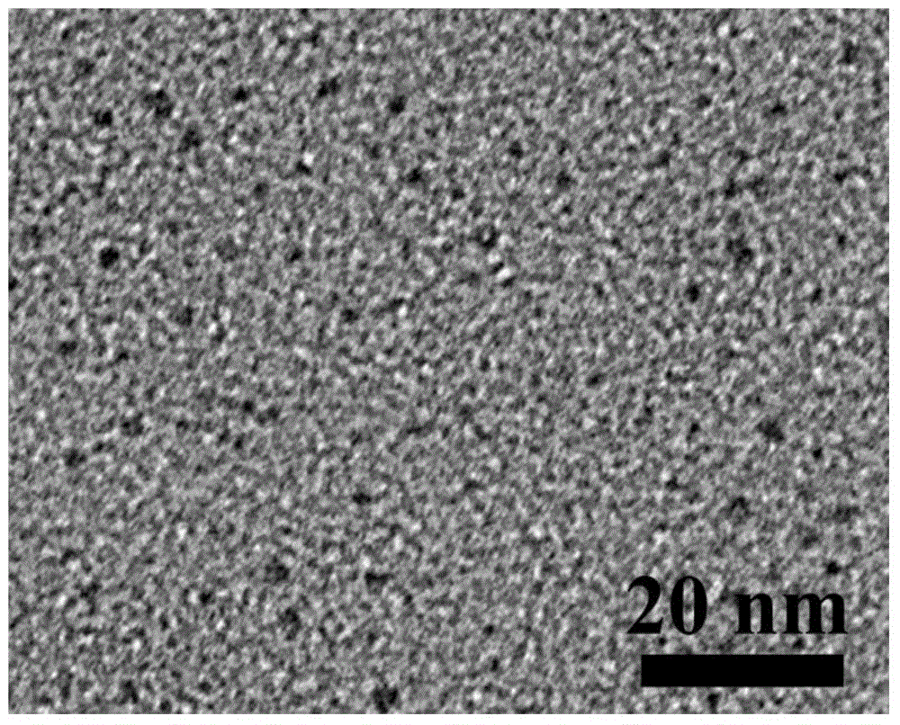
[0026] First, 3 g of citric acid and 3 g of urea were uniformly mixed in 10 mL of deionized water. The above solution was then placed in a 700 W microwave for 5 min, followed by heating at 60 °C in vacuo for 1 h. Centrifuge at 3000rpm for 20min to remove large particle impurities, and take out the upper layer liquid for later use.

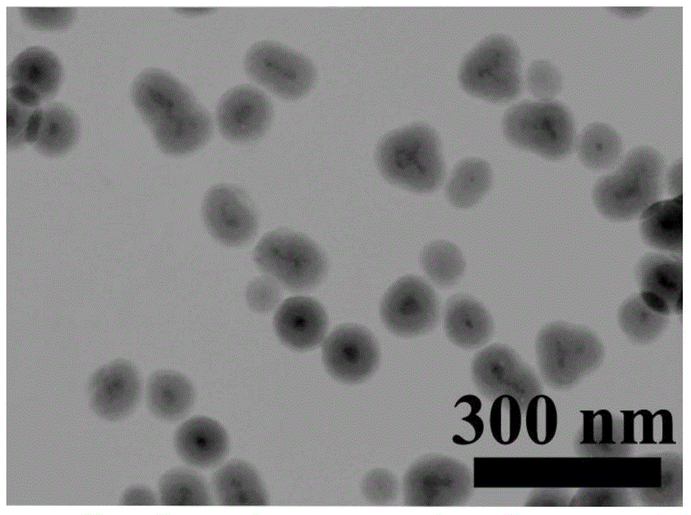
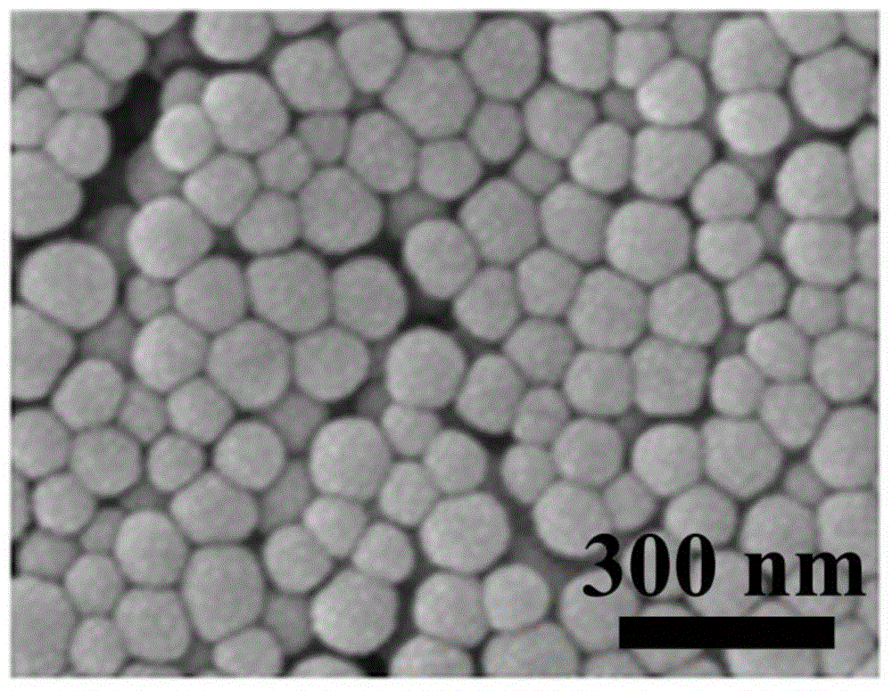
[0027] Take 350 μL of the above C-dots solution, add to 10mL PAAS aqueous solution (2gL -1 ), followed by adding 50 mL of ethanol and stirring for 30 min. Take the solution obtained in the above steps, add 180mL ethanol and 70mLNH 3 ·H 2 O (2M), after stirring evenly, add 20% TEOS solution (prepared with ethanol) several times, and react at 40°C for 2h. (First, add 100 μLTEOS, then add 100 μL after 40 minutes, and add 200 μL to the remaining 800 μLTEOS every 20 minutes) Finally, the obtained mixed solution is centrifuged, and the obtained solid is alternately washed several times with deionized water and absolute ethanol to obtain C-dotsSiO 2...

Embodiment 2

[0029] First, 6 g of citric acid and 8 g of urea were uniformly mixed in 15 mL of deionized water. The above solution was then placed in a microwave at 850W for 20min, followed by heating at 80°C in vacuum for 1.5h. Centrifuge at 3000rpm for 20min to remove large particle impurities, and take out the upper layer liquid for later use.

[0030] Take 500 μL of the above C-dots solution, add to 20mL PAAS aqueous solution (2gL -1 ), followed by adding 100mL of ethanol and stirring for 45min. Take the solution obtained in the above steps, add 100mL ethanol and 50mLNH 3 ·H 2 O (2M), after stirring evenly, add 20% TEOS solution (prepared with ethanol) several times, and react at 45°C for 2.5h. (First, add 250 μLTEOS, then add 250 μL after 40 minutes, and add 300 μL to the remaining 2000 μLTEOS every 20 minutes) Finally, the obtained mixed solution is centrifuged, and the obtained solid is alternately washed several times with deionized water and absolute ethanol to obtain C-dotsS...

Embodiment 3

[0032] First, 8 g of citric acid and 10 g of urea were uniformly mixed in 20 mL of deionized water. The above solution was then placed in a 1000 W microwave for 10 min, followed by heating at 100 °C in vacuum for 2 h. Centrifuge at 6000rpm for 20min to remove large particle impurities, and take out the upper layer liquid for later use.

[0033] Take 500 μL of the above C-dots solution, add to 20mL PAAS aqueous solution (2gL -1 ), followed by adding 100mL of ethanol and stirring for 60min. Take the solution obtained in the above steps, add 150mL ethanol and 100mL NH 3 ·H 2O (2M), after stirring evenly, add 25% TEOS solution (prepared with ethanol) several times, and react at 50°C for 3h. (First, add 400 μLTEOS, then add 400 μL after 40 minutes, and add 700 μL to the remaining 2800 μLTEOS every 20 minutes) Finally, the obtained mixed solution is centrifuged, and the obtained solid is alternately washed several times with deionized water and absolute ethanol to obtain C-dots...


PUM


Abstract

The invention provides a simple and green preparation method for monodisperse core-shell-structured C-dots@SiO2 fluorescence imaging nanoprobe. The method comprises the following steps: synthesizing a carbon quantum dot (C-dots) fluorescent nano-material with a uniform size, good dispersibility and high quantum efficiency by using a microwave method; and then coating C-dots in an ethanol-water system with ethyl orthosilicate (TEOS) as a silicon source so as to form C-dots@SiO2 nanoprobe suitable for biological imaging. Due to coating of SiO2, the prepared nanoprobe is easy to separate and wash and has stable physicochemical properties and optical properties, good biocompatibility and friendliness to the environment; moreover, surface modification of the nanoprobe can be easily carried out; and the nanoprobe is applicable to other biomedical clinical application fields, e.g., targeting imaging of cancer cells.

Description

technical field [0001] The invention belongs to the technical field of nanocomposite materials and their applications, in particular to a simple and green method for preparing monodisperse core-shell structure C-dotsSiO 2 Methods of fluorescent imaging nanoprobes and their application in in vivo imaging. Background technique [0002] Cancer is the most lethal disease in the world, so early detection and tracking of cancer cells is of great significance. There are many techniques for early cancer diagnosis. For example: CT is the most commonly used diagnosis and treatment method, which can clearly present the 3D structure of the tissue, but the vast majority of CT contrast agents used in clinical practice are iodide, and excessive application of iodide will cause great damage to the kidneys. The resolution of MR imaging is not so high. Since fluorescence imaging does not cause damage to the kidney, and has better imaging resolution at the cellular level. Therefore, the ap...

Claims


Application Information

Patent Timeline
Patent Type & Authority Applications(China)
IPC IPC(8): A61K49/00B82Y5/00
Inventor 王春刚吴晓彤苏忠民
Owner NORTHEAST NORMAL UNIVERSITY
Why Eureka
Try Eureka