Ultrasonic echo statistical parameter imaging system and method for thermal coagulation monitoring
An ultrasonic echo and imaging system technology, applied in the direction of ultrasonic/sonic/infrasonic Permian technology, ultrasonic/sonic/infrasonic image/data processing, ultrasonic/sonic/infrasonic diagnosis, etc., can solve the problem of coagulation initiation and coagulation Difficulties in bottom detection and monitoring imaging, etc., to achieve the effect of optimizing detection and monitoring imaging effects, enhancing the display of the lower part of solidification, and improving functions
- Summary
- Abstract
- Description
- Claims
- Application Information
AI Technical Summary
Problems solved by technology
Method used
Image
Examples
Embodiment 1
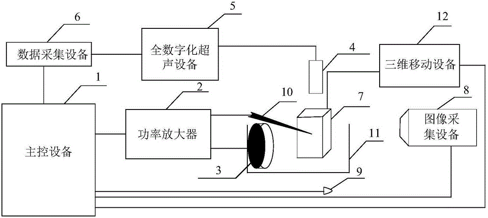
[0072] see Figure 4 , Coagulation initiation detection and ultrasound monitoring imaging during thermal ablation. In the early stage of thermal ablation therapy, the ultrasound echo statistical parametric imaging method was used to detect and monitor the onset of thermal coagulation.
[0073] In this embodiment, focused ultrasound is used for thermal ablation therapy. The sound power used is 85W, the duty cycle is 70%, and the total time is 4s. During the thermal ablation process, the image acquisition equipment is used to collect the dynamic change process of the damage in the transparent biological tissue phantom during the thermal ablation treatment with focused ultrasound. If shown in 4(a). At the same time, in the process of thermal ablation, the ultrasonic echo backscatter data of the target area in the process of thermal ablation treatment is collected synchronously with the all-digital ultrasound equipment. Use these collected ultrasound echo RF data to simultaneou...
Embodiment 2
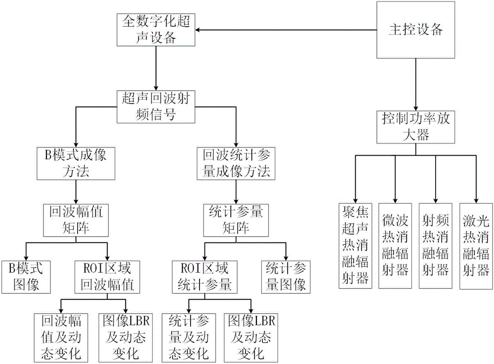
[0075] see Figure 5 , the dynamic changes and monitoring imaging of the statistical parameters of the lower part of the ultrasonic echo during thermal coagulation when the upper part of the hyperechoic area blocks the heat during the thermal ablation process. During the thermal ablation process, the ultrasonic echo amplitude and ultrasonic echo statistical parameters of multiple regions of the sample are detected synchronously. At the same time, the ultrasonic echo statistical parameter image and the ultrasonic B-mode image are constructed simultaneously.
[0076] In this embodiment, a microwave therapeutic apparatus is used for thermal ablation therapy. The parameters of microwave thermal ablation are: the ablation power is 40W, and the ablation time is 5min. During the thermal ablation process, the ultrasonic detection and imaging transducers are controlled by fully digital ultrasonic equipment to collect the ultrasonic echo radio frequency data of the thermal ablation tar...
Embodiment 3
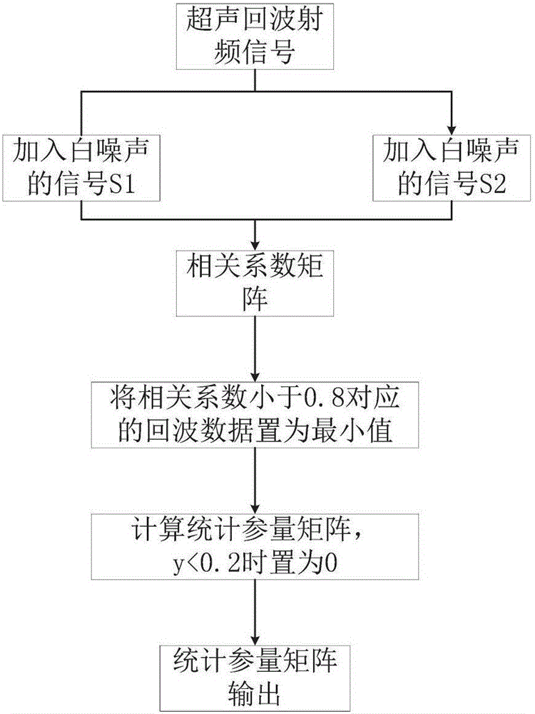
[0078] see Figure 6 , Monitoring imaging of thermal coagulation based on ultrasonic echo-statistical parameters increasing the ratio of thermally coagulated microbubbles. During the thermal ablation process, the ultrasonic echo data and ultrasonic echo statistical parameter data of the coagulation area and the microbubble area are respectively extracted, the average amplitude of the ROI area is calculated, and the LBR value and its dynamic changes are calculated according to the formula. Echo statistical parametric images and B-mode ultrasound images.
[0079] In this embodiment, focused ultrasound is used for thermal ablation treatment, the sound power is 140W, and continuous radiation is 32s. During the thermal ablation treatment, a fully digital ultrasound device is used to synchronously collect ultrasonic echo radio frequency data of the target area during the thermal ablation process. Use these RF data to simultaneously construct B-mode images during thermal ablation ...
PUM
 Abstract
Description
Claims
Application Information
 - R&D
- Intellectual Property
- Life Sciences
- Materials
- Tech Scout
- Unparalleled Data Quality
- Higher Quality Content
- 60% Fewer Hallucinations
Browse by: Latest US Patents, China's latest patents, Technical Efficacy Thesaurus, Application Domain, Technology Topic, Popular Technical Reports.
© 2025 PatSnap. All rights reserved.Legal|Privacy policy|Modern Slavery Act Transparency Statement|Sitemap|About US| Contact US: help@patsnap.com


