
A car-t cytotoxicity indicator carrier

A cytotoxic and cellular technology, applied in the direction of virus/bacteriophage, retroRNA virus, using vectors to introduce foreign genetic material, etc., can solve the research and application development constraints of CAR-T cell therapy technology, long detection time, lack of CAR-T T cell toxicity detection method R&D and application of CAR-T cell therapy technology to achieve excellent production prospects, shortened time, excellent flexibility and specificity

Active Publication Date: 2017-12-22
济南宜明医疗科技有限公司
View PDF3 Cites 0 Cited by
  • Summary
  • Abstract
  • Description
  • Claims
  • Application Information

AI Technical Summary

Problems solved by technology

[0003] In medical research and practical application, the indication or characterization of CAR-T cell toxicity is an inevitable and important problem. If this problem cannot be better solved, the research and application development of CAR-T cell therapy technology will be affected. severe constraints
However, the currently widely used method is to use CAR-T to kill target cells cultured in vitro, and to detect its effect through indirect measurement methods, so as to draw experimental conclusions; in the currently commonly used CAR-T cytotoxicity assay methods, in After CAR-T cells and target cells are mixed, it usually takes more than two days of culture (such as MTT colorimetric method, etc.), so the required detection time is relatively long
The lack of direct and efficient CAR-T cytotoxicity detection methods has become a major obstacle to the development and application of CAR-T cell therapy technology

Method used


Image

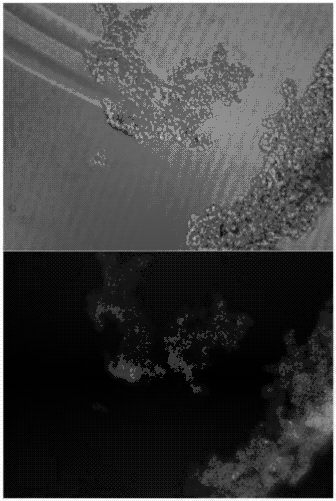
  • A car-t cytotoxicity indicator carrier
  • A car-t cytotoxicity indicator carrier
  • A car-t cytotoxicity indicator carrier

Examples


Embodiment 1

[0033] Example 1 Construction of CAR-T cytotoxicity indicator vector

[0034] (1) Synthesis of CD19-P2A- Luciferase sequence, and introduce Asis I and MluI restriction sites in the upstream and downstream respectively;

[0035] (2) Enzyme digestion of Plent-EF1α-Puro-CMV vector and CD19-P2A containing Asis I and MluI restriction sites obtained in step (1) Luciferase sequence, enzyme digestion system is as follows:

[0036]

[0037]

[0038] After adding and mixing the sample, place it at 37°C for enzyme digestion for 3 hours. After the reaction, use 1% agarose gel electrophoresis to detect the digested CD19-P2A-Nanoluc Luciferase sequence and Plent-EF1α-Puro-CMV vector. And use the DNA gel recovery kit to recover the digested CD19-P2A- Luciferase sequence and Plent-EF1α-Puro-CMV vector;

[0039] (3) CD19-P2A- The Luciferase sequence and the Plent-EF1α-Puro-CMV vector were ligated at 22°C for 2 hours, and the ligation system was as follows,

[0040]

[0041] ...

Embodiment 2

[0042] Embodiment 2 packaging lentivirus

[0043] (1) Transfer HEK293T cells whose cell growth is more than 90% of the maximum load capacity of a 10cm culture dish to a 10cm cell culture dish at a ratio of 1:3 (the number of cells per dish is about 2.5×10 6 ), placed in a 37°C, 5% CO2 incubator for 24 hours; obtain the cells to be transfected; the ratio of 1:3 refers to the cells in one 10cm culture dish, which are averagely passaged to three same culture dishes;

[0044] (2) Change the medium before transfection, and replace 5 ml of DMEM medium containing 10% FBS with the cells to be transfected obtained in step (1) with an electric pipette to obtain cells before transfection;

[0045] Prepare the transfection reagent as follows:

[0046]

[0047]

[0048] Prepare reagents 1 and 2 respectively, and place them at room temperature for 5-10 minutes; mix reagents 1 and 2 evenly, and place them at room temperature for 15-30 minutes to obtain reagent 3; add reagent 3 dropwis...

Embodiment 3

[0049] Example 3 Preparation of CAR-T cytotoxicity indicator cells

[0050] Infection experiments were carried out according to conventional methods known to those skilled in the art, and the infection steps are briefly described as follows:

[0051] (1) Lentivirus infection

[0052] In a 6-well plate, use the co-infection reagent ADV-HR and the lentiviral particles obtained in Example 2 to infect K562 cells, calculate the amount of lentiviral particles according to the multiplicity of infection (MOI=10), and use DMEM medium every other day during the infection process. Cells were exchanged to maintain the cell concentration at 0.5-2×10 6 / mL.

[0053] (2) Resistance screening

[0054] 72 hours after infection, start resistance screening:

[0055] Puromycin at a concentration of 2 μg / mL was selected for 14 days to obtain polyclonal cell lines with puromycin resistance, and monoclonal cell lines were obtained by further screening with puromycin at a concentration of 2 μg / mL...


PUM


Abstract

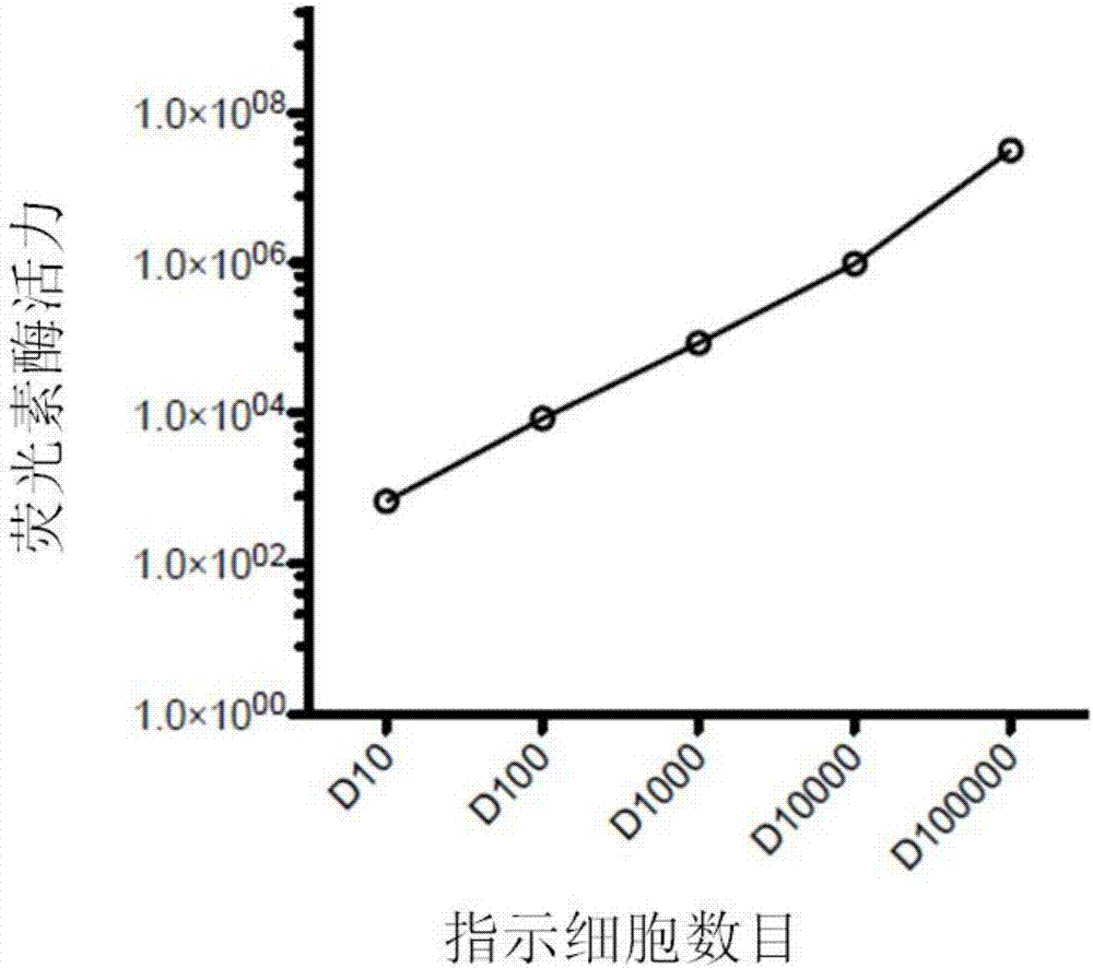
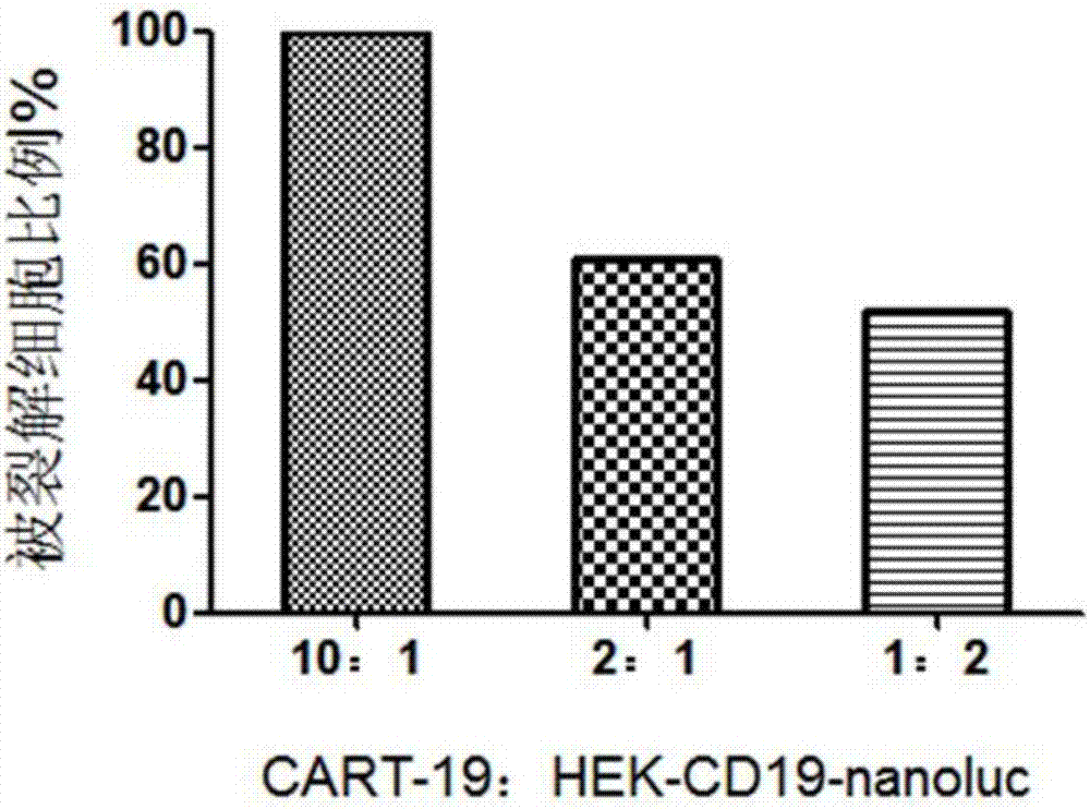
The invention belongs to the field of cellular immunotherapy and particularly relates to a CAR-T cytotoxicity indicating vector. The cytotoxicity indicating vector is a recombined lentiviral vector and is composed of an original lentiviral vector and an insertion sequence. The insertion sequence is composed of a tumor-correlation target antigen coding gene, a connection peptide coding gene and a luciferase coding gene from the 5' terminal to the 3'terminal in sequence. After CAR-T cells and CAR-T cytotoxicity indicating cells are mixed, a tumor-correlation target antigen expressed by the indicating cells can perform target guiding on CAR-T for combination, the indicating cells are split, and luciferase is released. Due to the fact that split cells are located in supernatant and the un-split cells are located in precipitate, the number of spit cells and the number of un-split cells can be worked out according to the relation between the indicating cell number and the luciferase activity value by detecting the activity value of luciferase in the supernatant and the activity value of luciferase in the precipitate, the proportion of the split cells is calculated, and the CAR-T cytotoxicity is indicated.

Description

technical field [0001] The invention belongs to the field of cellular immunotherapy, and in particular relates to a CAR-T cytotoxicity indicator carrier. Background technique [0002] CAR-T cells refer to T cells capable of expressing chimeric antigen receptor (CAR-T), which consists of an extracellular antigen-binding domain, a transmembrane domain, and an intracellular signaling domain. CAR-T cell therapy technology refers to the recombinant plasmid obtained by genetic recombination of the above-mentioned CAR structure in vitro and then introduced into the patient's T cells in vitro, so as to obtain CAR-T cells capable of expressing the above-mentioned chimeric antigen receptor. Then, after purification and large-scale expansion in vitro, the above-mentioned CAR-T cells are infused back into the patient to perform the function of killing tumor cells. The advantages of CAR-T therapy are: first, its combination with tumor antigens does not need to rely on the presentation o...

Claims


Application Information

Patent Timeline
Patent Type & Authority Patents(China)
IPC IPC(8): C12N15/867C12N5/10C12Q1/66C12Q1/06
CPCC07K14/70503C07K14/7051C07K14/70575C07K2319/61C12N15/86C12N2740/15043C12Q1/66
Inventor 孙秀莲李欣
Owner 济南宜明医疗科技有限公司
Learn More