Method for inducing and differentiating precursor cells of human skin source into corneal endothelial cells

A technology of precursor cells and corneal endothelium, applied in the field of tissue engineering and ophthalmic repair, can solve the problems of poor histocompatibility, lack of expanded functional corneal endothelial cells, lack of animal experiments, etc., to achieve immunogenicity and immune response Weak sex, suitable for clinical experiment and application, the effect of high induction differentiation efficiency

Active Publication Date: 2017-01-04
吴欣怡
View PDF1 Cites 3 Cited by
  • Summary
  • Abstract
  • Description
  • Claims
  • Application Information

AI Technical Summary

Problems solved by technology

Early researchers tried to use adipose mesothelial cells and umbilical cord blood mesenchymal stem cells to replace non-proliferative corneal endothelial cells, or to induce differentiation of embryonic stem cells, corneal stromal stem cells, bone marrow endothelial progenitor cells, and neural crest cells into corneal endothelial-like cells. However, these methods have certain problems, such as immune rejection, ethical issues, poor histocompatibility, lack of animal experiments, etc.
At present, there is still no method to achieve large-scale expansion of functional corneal endothelial cells in vitro

Method used


Image

  • Method for inducing and differentiating precursor cells of human skin source into corneal endothelial cells
  • Method for inducing and differentiating precursor cells of human skin source into corneal endothelial cells
  • Method for inducing and differentiating precursor cells of human skin source into corneal endothelial cells

Examples


Embodiment 1

[0047] Example 1 Inducing the differentiation of human skin-derived precursor cells into corneal endothelial-like cells

[0048] 1. Cultivation of SKPs precursor cells derived from human skin:

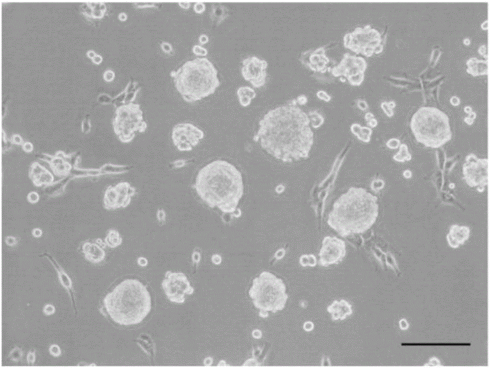
[0049] Rinse and disinfect the skin tissue with penicillin-streptomycin, cut into 1mm×2mm tissue pieces, digest with Dispase enzyme at 4°C for 12-24h, remove the epidermis to obtain the dermis, digest with collagenase for 2-3h, and use fetal bovine serum Neutralize with DMEM, dissociate the cells, filter them with a cell mesh, inoculate the cells in a culture flask, add SKPs culture medium, and place at 37°C, 5% CO 2 Cultured in an incubator. After about 2-3 weeks, spherical suspension SKPs are formed, which are used to induce differentiation when passed to 2-4 passages. The culture observation results of SKPs cells are shown in figure 1 shown.

[0050] SKPs culture medium is DMEM / F12=3:1 basal medium, supplemented with 2% B27, 40ng / ml bFGF, 20ng / ml EGF, 1% green chain double antib...

Embodiment 2

[0057] Example 2 Identification of corneal endothelial-like cells and corneal repair experiment

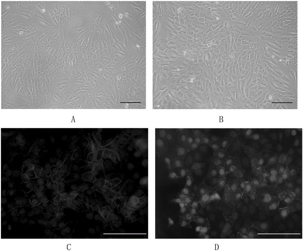
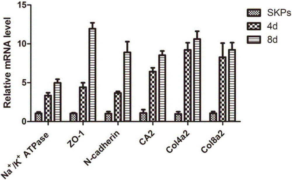
[0058] After 4 days of induction, part of the cells transformed into polygonal shapes, which gradually increased with time. After 8 days, polygonal cells accounted for the majority, forming tight connections with each other in a monolayer mosaic-like arrangement. Optical microscope observation, immunofluorescence, real-time quantitative PCR, Western Blotting confirmed that the induced cells had similar morphology and marker expression to human keratocytes. Corneal endothelial-like cells can be stably passaged for 3-4 passages, and can still maintain their morphology and the expression of various markers after passage. The optical microscope effect picture of SKPs induced to differentiate into corneal endothelial-like cells can be found in figure 2 shown. RT-PCR results and Westernblotting test results respectively refer to image 3 , Figure 4 .

[0059] Rabbit corneal endot...


PUM


Abstract

The invention discloses a method for inducing and differentiating precursor cells of a human skin source into corneal endothelial cells. The precursor cells of the human skin source are used and cultured together with the corneal endothelial cells B4G12, corneal endothelial cells theoretically approaching corneal endothelial cells of a normal person are successfully induced, the obtained corneal endothelial cells are applied to a corneal endothelial decompensation animal model, the corneal endothelium of an animal is successfully repaired, and the method has important clinical application prospects.

Description

technical field [0001] The invention relates to a method for inducing differentiation of human skin-derived precursor cells into corneal endothelial-like cells, and belongs to the technical fields of tissue engineering and ophthalmic repair. Background technique [0002] In the five-layer structure of the cornea, the innermost endothelial layer is very important for maintaining corneal transparency and normal physiological functions. Human corneal endothelial cells (HCECs) through the pump function and barrier function, Regulates the transparency of the cornea. However, due to the lack of proliferation ability of adult corneal endothelial cells, damaged corneal endothelial cells mainly rely on the expansion and migration of peripheral cells in the damaged area for repair. When the cell density is less than 500-800 / mm 2 At this time, it will cause decompensation of the corneal endothelium, resulting in continuous corneal edema and loss of transparency. Infection, inflammati...

Claims


Application Information

Patent Timeline
Patent Type & Authority Applications(China)
IPC IPC(8): C12N5/071A61L27/38
CPCA61L27/3808C12N5/0621A61L2430/16C12N2506/09A61L27/3839C12N2502/09A61L27/3604C12N2500/30C12N2500/90C12N2501/115C12N2509/00
Inventor 吴欣怡申琳
Owner 吴欣怡
Why Eureka
Try Eureka