

Method of using three dimensional reconstruction imaging technology to analyze structure of root tip cells of arabidopis thaliana
A technology of technical analysis and cell structure, applied in the field of cell structure analysis, can solve problems such as occlusion and inaccurate results, and achieve the effect of improving resolution, ensuring fineness, and reducing shooting steps.
- Summary
- Abstract
- Description
- Claims
- Application Information
AI Technical Summary
Problems solved by technology
Method used
Image

Examples
Embodiment Construction
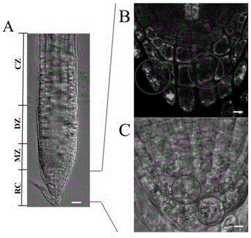
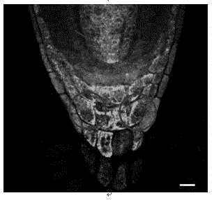
[0027] 1. Experimental materials
[0028] 1.1 Plant material
[0029] Arabidopsis Columbia Type 0 Gifted by Professor Han Shengcheng, School of Life Sciences, Beijing Normal University
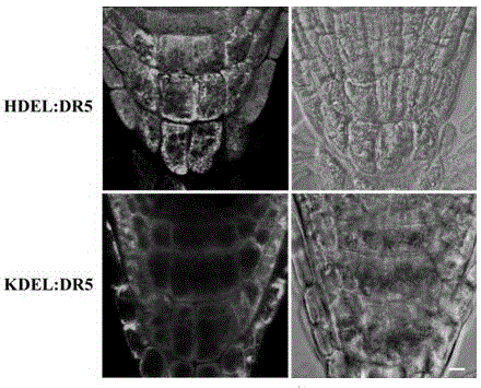
[0030] Arabidopsis DR5: HDEL Gift from Professor Lin Jinxing, Institute of Botany, Chinese Academy of Sciences
[0031] Arabidopsis DR5: KDEL Gift from Professor Le Jie, Institute of Botany, Chinese Academy of Sciences
[0032] 1.3 Main reagents and consumables
[0033] MS Medium PhytoTech
[0034] Propidium iodide Sigma
[0035] OCT frozen section embedding medium Leica
[0036] Triton X-100 Beijing Dingguochangsheng Biotechnology Co., Ltd.
[0037] 0.22um PVDF filter Millipore
[0038] 1.4 Main instruments and equipment
[0039] CM3050S Cryostat Leica
[0040] Upright fluorescence microscope ImagerA1 Carl Zeiss
[0041] Confocal Laser Microscope LSM 510 Carl Zeiss
[0042] Confocal Laser Microscope-FV300 Olympus
[0043] Intelligent light incubator-GZH-268B Hangzhou Huier Instrum...
PUM
 Abstract
Description
Claims
Application Information