Construction method and application of human normal vaginal epithelium 3D (Three Dimensional) differentiation culture model

A construction method and a technology for differentiation medium, which are applied in the field of construction of a 3D differentiation culture model of human normal vaginal epithelium, and can solve the problems of insignificant effect and inability to further study the pathogenic mechanism of infection.

Active Publication Date: 2017-05-10
深圳涌泰生物科技有限公司
View PDF2 Cites 13 Cited by
  • Summary
  • Abstract
  • Description
  • Claims
  • Application Information

AI Technical Summary

Problems solved by technology

Therefore, there are always obvious limitations in using conventional two-dimensional cell culture models to study the infection mechanism of sexually transmitted pathogens
On the other hand, most studies in recent decades have used animal models to study viral HSV-2 vaccines. Due to the species differences between animals and humans, the effect of HSV-2 vaccines developed through animal models on humans is not obvious , so these HSV-2 vaccines at the level of animal research are rarely used clinically
At the same time, the use of animal models cannot further study the pathogenic mechanism of microscopic HSV-2 infection

Method used


Image

  • Construction method and application of human normal vaginal epithelium 3D (Three Dimensional) differentiation culture model
  • Construction method and application of human normal vaginal epithelium 3D (Three Dimensional) differentiation culture model
  • Construction method and application of human normal vaginal epithelium 3D (Three Dimensional) differentiation culture model

Examples


Embodiment 1

[0052] [Example 1] Primary isolation and culture of human normal vaginal epithelial cells

[0053] (1) With the informed consent of the patient or the patient guardian, collect the paracancerous normal tissue samples from surgically resected patients with vaginal cancer.

[0054] (2) Preparation of digestive solution: primary epithelial cell growth medium containing 0.2 mg / mL of collagenase and dispase; wherein, the components of primary epithelial cell growth medium (2D medium) include: DMEM and Ham's F-12NUTRIENT MIX mixed medium at a volume ratio of 3:1, while adding 5% fetal bovine serum, 2nM triiodothyronine (triiodothyronine), 0.5% insulin iron selenium transfer protein (insulin-transferrin-selenium) reagent, 5μg / ml transferrin (transferrin), 10ng / mL epidermal growth factor, 0.4μg / mL hydrocortisone, 40μg / mL gentamicin, 50nM calpeptin, 40ng / ml recombinant human IL-1RA, and 3μg / ml recombinant Human R-Spondin-1.

[0055] (3) Wash the isolated tissue sample once with 95-1...

Embodiment 2

[0063] [Example 2] subculture of human normal vaginal epithelial cells

[0064] (1) When the human normal vaginal epithelial cells cultured in T25 or T75 culture flasks proliferate to 70-90% abundance, wash the cells three times with 1×PBS (0.01M, pH 7.4), and then wash them with 0.05% (mass Volume ratio) Trypsin-EDTA digested monolayer cells for 2-5 minutes.

[0065] (2) Add 10mL DMEM to neutralize the digestion reaction for 1-2 minutes.

[0066] (3) Centrifuge at 1000rmp for 5 minutes, discard the supernatant.

[0067] (4) Resuspend the cell pellet in 2D medium at a ratio of 1:2, 1:3, 1:4 or 1:5, inoculate it in a culture flask, and culture it at 37°C, 5% CO 2 Human normal vaginal epithelial cells were obtained.

[0068] (5) If necessary, 1×10 6 Epithelial cells were resuspended in 1-2 mL of cell freezing medium (90% fetal bovine serum and 10% DMSO, v / v), and stored in liquid nitrogen for future use.

Embodiment 3

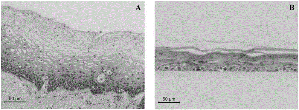
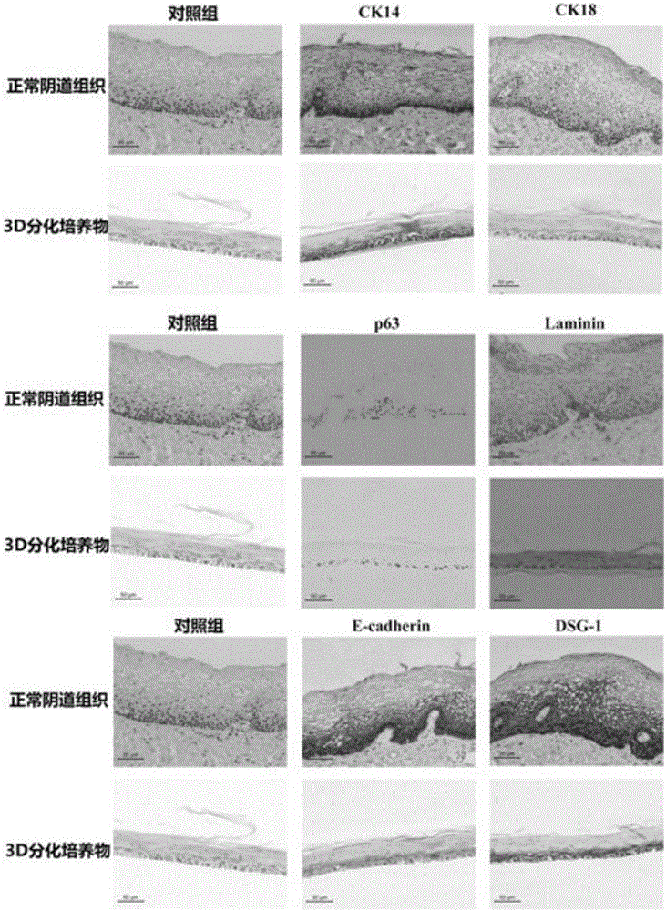
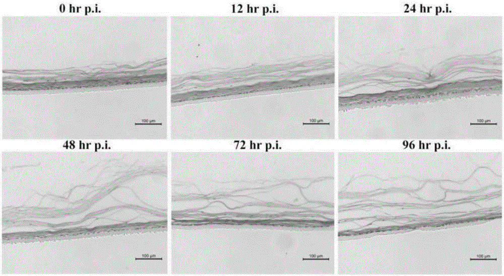
[0069] [Example 3] Air-liquid 3D culture of human normal vaginal epithelium

[0070] (1) Put 0.4μm Millicell PCF insert (12mm size, Millipore) into a six-well plate, and put up to three inserts in each well.

[0071] (2) Resuspend 5x10 with 400μl 2D medium 5 Individual human vaginal epithelial cells were seeded into each insert.

[0072] (3) Add 2ml of 2D medium to the inside of each well plate, that is, to the periphery of the insert.

[0073] (4) Put the six-well plate with the insert in a humid heat incubator at 37°C, 5% CO 2 The incubation period was 48 hours.

[0074] (5) Replace the medium inside and outside the insert with a 3D differentiation medium.

[0075] The preparation of 3D differentiation medium was: DMEM and F12 were mixed at a volume ratio of 1:1, and 0.87 μM insulin (Sigma-Aldrich I6634), 0.125 μM transferrin (Sigma-Aldrich T0665), 0.1 μM hydrocortisone were added at the same time. Pine (Sigma-Aldrich H0396), 0.01 μM triiodothyronine (Sigma-Aldrich T639...


PUM

PropertyMeasurementUnit
thicknessaaaaaaaaaa

Abstract

The invention belongs to the field of biomedicines and discloses a construction method and application of a human normal vaginal epithelium 3D (Three Dimensional) differentiation culture model. A 2D (Two Dimensional) growth culture medium is provided and normal vaginal epithelial cells separated and cultured from normal tissues beside female vaginal cancer are obtained; any exogenous gene is not introduced and the normal vaginal epithelial cells have a physiological function of normal differentiation. The method for constructing the human normal differentiation vaginal epithelium 3D model comprises the following steps: re-suspending single cells by the 2D culture medium and inoculating the single cells into a gas-liquid culture device; replacing the growth culture medium with a differentiation culture medium and culturing for 14 to 21 days; after completely differentiating 3D gas-liquid culture of the human vaginal epithelial cells, inoculating HSV-2 (Herpes Simplex Virus-2) virus liquid to obtain an HSV-2 virus infection 3D model. The two types of 3D models can be used for physiological studies and drug toxicity safety evaluation of human normal reproductive tract epithelium, researches of pathogenic mechanisms of HSV-2 virus infected diseases and sexual transmission pathogen infected diseases and researches and development of anti-viruses medicines.

Description

technical field [0001] The invention belongs to the field of cell biology, and relates to a method for constructing a human normal vaginal epithelial 3D differentiation culture model and its application. Background technique [0002] Vital organs of the human body are composed of organ-specifically differentiated epithelial cells. These specifically differentiated epithelial cells are directly related to the specific functions of different organs. The closest to the lumen of the genital tract is the epithelial tissue layer on the mucous membrane of the genital tract. The epithelial layer is exposed to various pathogens in the lumen of the genital tract and separates the pathogens in the genital tract from the reproductive system. Therefore, the reproductive tract epithelium is the first natural physical barrier for the body to resist external pathogens. Once these vital organs become diseased or degenerate, human health will be threatened, because these vital organs are di...

Claims


Application Information

Patent Timeline
Patent Type & Authority Applications(China)
IPC IPC(8): C12N5/071C12Q1/02
CPCC12N5/0625C12N5/0682C12N2500/22C12N2500/25C12N2500/38C12N2500/46C12N2501/11C12N2501/2301C12N2501/39C12N2501/395C12N2501/81G01N33/5014G01N33/5044
Inventor 李晖叶立娜朱雅琪丘建斌吴小婷
Owner 深圳涌泰生物科技有限公司
Why Eureka
Try Eureka