Preparation method and detection method of carcino-embryonic antigen electrochemical immunosensor

An immunosensor and carcinoembryonic antigen technology, applied in the field of immunosensors, can solve the problems of unstable electrochemical response, complex detection steps, wrong detection results, etc., to eliminate the interference of dissolved oxygen, high selective permeability, and low cost. Effect

Inactive Publication Date: 2017-05-31
CHONGQING UNIV OF ARTS & SCI
View PDF3 Cites 2 Cited by
  • Summary
  • Abstract
  • Description
  • Claims
  • Application Information

AI Technical Summary

Problems solved by technology

Enzyme biosensors detect the H produced by enzymatic reactions 2 o 2 , but this type of enzyme biosensor will be severely affected by dissolved oxygen, resulting in unstable electrochemical response
Commonly used bioelectrochemical sensors require relatively complicated detection steps and need to be thoroughly cleaned before use, which is very easy to cause errors in the detection results

Method used


Image

  • Preparation method and detection method of carcino-embryonic antigen electrochemical immunosensor
  • Preparation method and detection method of carcino-embryonic antigen electrochemical immunosensor
  • Preparation method and detection method of carcino-embryonic antigen electrochemical immunosensor

Examples


Embodiment 1

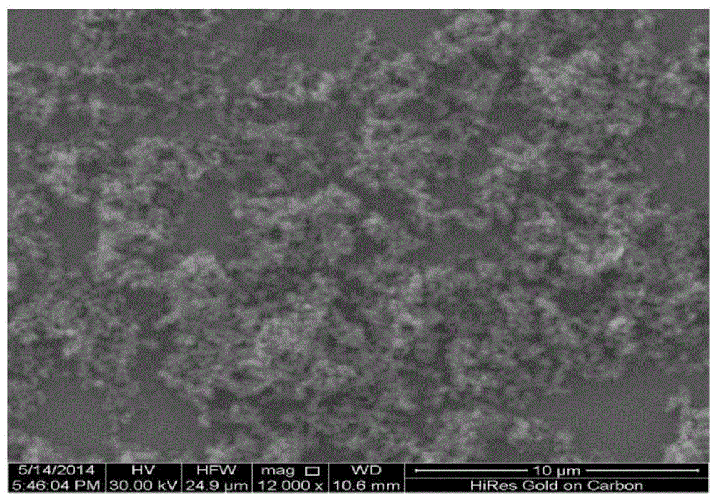
[0037] 1. Preparation of poly-o-phenylenediamine nanospheres (POPD)

[0038] Add 200μL of 0.1M o-phenylenediamine to 3mL of water, inject 30ml / min of ozone into the solution and keep stirring. After stirring for 5h at room temperature, centrifuge the reaction product under a gravity acceleration of 7000G for 5min to separate the precipitate That is, poly-o-phenylenediamine nanospheres.

[0039] 2. Preparation of immunosensor

[0040] Step 1: Use 0.3 μm and 0.05 μm Al on the glassy carbon electrodes, respectively 2 o 3 polishing;

[0041] Step 2: First use 1:1 HNO 3 After ultrasonic cleaning for 5 min, ultrasonic cleaning with absolute ethanol for 5 min, and finally pure water ultrasonic cleaning for 5 min; at a deposition potential of 1.75 V, in 0.1M PBS with pH=5.0, linear scan stripping voltammetry was used to deposit for 300 s, and the obtained Treated glassy carbon electrode;

[0042] Step 3: Perform cyclic voltammetry curves on the treated glassy carbon electrode in...

Embodiment 2

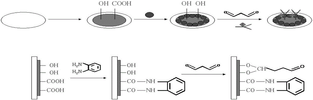
[0055] Example 1 was repeated according to the same steps described above, but the stirring time was 3 hours after ozone was introduced into the preparation of poly-ortho-phenylenediamine. The cleaning agent in the preparation of the immunosensor is 1:1 nitric acid, toluene, and deionized water; immersion in 20mM glutaraldehyde solution for 24h; the glutaraldehyde-modified glassy carbon electrode was obtained on the homemade poly-o-phenylenediamine nanomicro Immerse in the mixture of ball solution for 10h. The dried immunosensor was soaked in PBS solution containing 0.04% BSA at 4° C. for 3 h.

[0056] Finally, an electrochemical immunosensor for detecting carcinoembryonic antigen was prepared.

Embodiment 3

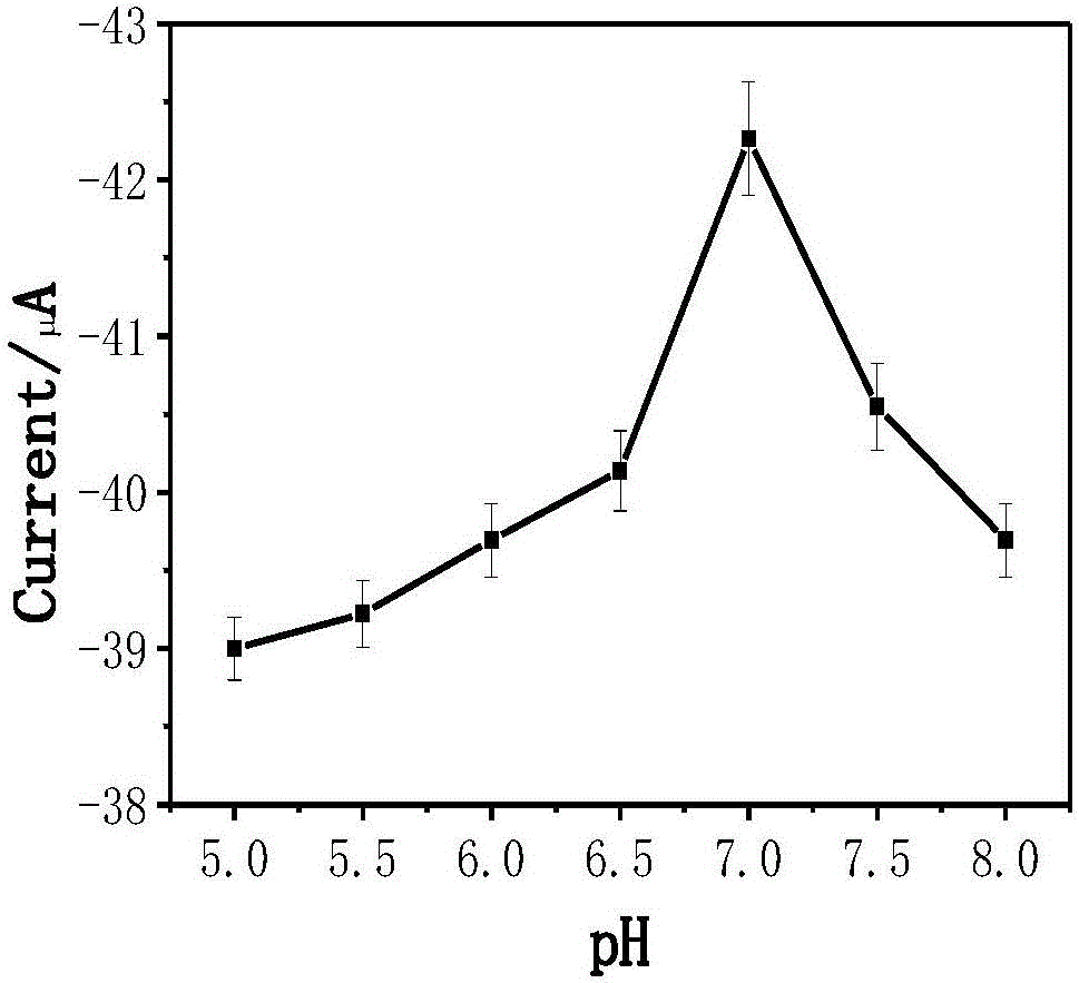
[0058] Example 1 was performed following the same procedure as described, but with the pH of the anaerobic PBS solution = 5.0.


PUM


Abstract

The invention belongs to the field of immunosensor, and more specifically discloses a preparation method and a detection method of a carcino-embryonic antigen (CEA) electrochemical immunosensor. The preparation method comprises following steps: poly o-phenylenediamine nanoparticles are dispersed in water via ultrasonic oscillation, and an obtained poly o-phenylenediamine nanoparticle solution is stored at room temperature; a glassy carbon electrode is modified with glutaraldehyde, and the glassy carbon electrode is immersed in a mixed solution composed of a HRP labeled CEA antibody solution and the poly o-phenylenediamine nanoparticle solution so as to obtain a poly o-phenylenediamine nanoparticle / HRP labeled CEA antibody modified electrode; solutions with different concentrations are prepared from CEA standard samples, and are taken as test solutions; an analyte is subjected to differential pulse voltammetry scanning in a H2O2 and oxygen-free PBS mixed solution; electrochemical reaction is carried out so as to establish the quantitative relationship of electrochemical current strength with the CEA solution concentration. The carcino-embryonic antigen electrochemical immunosensor is high in sensitivity; results are accurate and reliable; cost is low; detection is rapid; the carcino-embryonic antigen electrochemical immunosensor is convenient to use; and the preparation process is extremely simple.

Description

technical field [0001] The invention relates to the field of immunosensors, in particular to a preparation method and detection method of a carcinoembryonic antigen electrochemical immunosensor. Background technique [0002] Carcinoembryonic antigen (CEA) is a glycoprotein with extremely large molecular weight, which is often used as a tumor marker. The detection of CEA concentration can not only be used in the diagnosis of tumors in the gastrointestinal tract and respiratory tract, but also can indicate the nature of the tumor by detecting the content and amount of carcinoembryonic antigen in the human body, so as to diagnose the tumor, analyze the course of the disease and guide clinical treatment. play an auxiliary role. Therefore, the determination of CEA concentration has great significance for clinical tumor diagnosis. The most common analytical methods for quantitative detection of CEA include fluorescent immunoassay, enzyme-linked immunosorbent assay, electrochemic...

Claims


Application Information

Patent Timeline
IPC IPC(8): G01N27/327
CPCG01N27/3278G01N27/3271G01N27/3277
Inventor 田亮亮李璐陈贤闫兴武刘碧桃程江杨鑫
Owner CHONGQING UNIV OF ARTS & SCI
Why Eureka
Try Eureka