Fibroin protein fiber scaffold and preparation method thereof

A fibrous scaffold and silk fibroin technology, applied in medical science, prostheses, etc., can solve the problems of complex processing, unfavorable cell and tissue growth, and limited tissue growth in electrospinning technology, and achieve easy batch processing and excellent Mechanical properties, the effect of reducing purification steps and time

Active Publication Date: 2017-07-11
上海丝波敦生物科技有限公司
View PDF5 Cites 7 Cited by
  • Summary
  • Abstract
  • Description
  • Claims
  • Application Information

AI Technical Summary

Problems solved by technology

[0004] At present, there are many methods for preparing porous silk fibroin scaffolds, including freeze-drying, salting-out method, gas foaming method, three-dimensional printing, etc. However, these methods still have some insurmountable shortcomings.
For example, the freeze-drying method is easy to form a sheet structure, which is not conducive to the growth of cells and tissues. Although the prior art has reported a method of repeated membrane dissolution to control the self-assembly of silk fibroin to form a nanofibrous structure, and then form a porous scaffold, the efficiency is low. Poor reproducibility, and the existence of pore wall structure directly restricts cell migration and interaction, and tissue growth is thus limited
The fiber scaffold prepared by electrospinning technology is considered to be an ideal scaffold structure for tissue engineering; however, the electrospinning technology is complex in processing, low in yield, and the structure of the electrospun fiber membrane is dense, which is not conducive to the growth of cells and tissues

Method used


Image

  • Fibroin protein fiber scaffold and preparation method thereof
  • Fibroin protein fiber scaffold and preparation method thereof
  • Fibroin protein fiber scaffold and preparation method thereof

Examples


Embodiment 1

[0031] (1) Natural mulberry silk was degummed by boiling 0.05wt% sodium carbonate solution for 30 minutes, and obtained silk fiber after repeating 3 times; the silk fiber was dispersed in 88% formic acid solvent, and the concentration of the obtained silk fiber dispersion was 2% ;

[0032] (2) Inject the silk fiber dispersion liquid into the mold for freezing treatment (temperature -20°C) to obtain silk fiber frozen body;

[0033] (3) immerse the frozen silk fiber body in step (2) in methanol to remove formic acid, and then soak and wash with deionized water to obtain a wet silk fibroin fiber scaffold;

[0034] (4) Freezing the wet silk fibroin fiber scaffold obtained in step (3) at -20°C, and then freeze-drying at -40°C to obtain a silk fibroin scaffold.

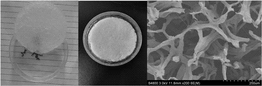
[0035] attached figure 1 It is the photos of the wet (left) and dry (middle) silk fibrous scaffolds obtained above and the scanning electron microscope picture (right). It can be seen from the figure that the interior of ...

Embodiment 2

[0037] (1) Natural mulberry silk was degummed by boiling 0.5wt% sodium carbonate solution for 30 minutes, and the silk fiber was obtained after repeating 3 times; the silk fiber was dispersed in 98% formic acid solvent, and the concentration of the obtained silk fiber dispersion was 3% ;

[0038] (2) Inject the silk fiber dispersion liquid into the mold for freezing treatment (temperature -10°C) to obtain silk fiber frozen body;

[0039] (3) immerse the frozen silk fiber body in step (2) in ethanol to remove formic acid, and then soak and wash with deionized water to obtain a wet silk fibroin fiber scaffold;

[0040] (4) The wet silk fibroin fiber scaffold was frozen at -10°C, and then freeze-dried at -20°C to obtain the silk fibroin scaffold.

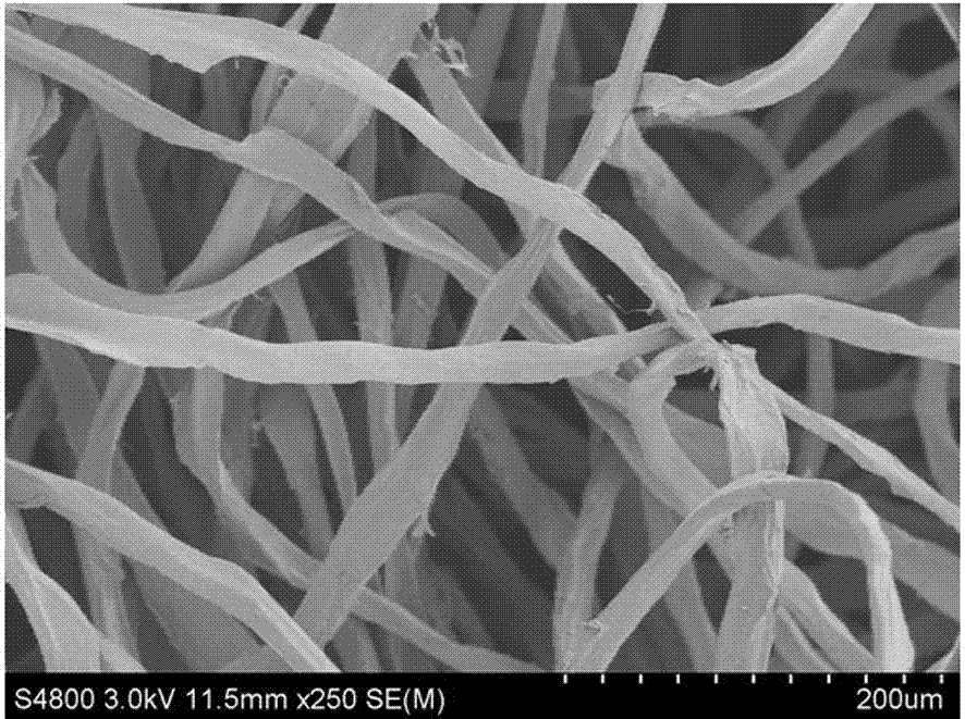
[0041] attached figure 2 The photo (left) and scanning electron microscope image (right) of the silk fiber scaffold obtained above; it can be seen from the figure that the interior of the scaffold is mainly composed of fibers, and s...

Embodiment 3

[0043] (1) Natural tussah silk was degummed by boiling 0.05wt% sodium carbonate solution for 30 minutes, and obtained silk fiber after repeating 3 times; the silk fiber was dispersed in 98% formic acid solvent, and the concentration of silk fiber dispersion obtained was 5% ;

[0044] (2) Inject the silk fiber dispersion liquid into the mold for freezing treatment (temperature -30°C) to obtain silk fiber frozen body;

[0045] (3) Immerse the frozen silk fiber body in step (2) in propanol to remove formic acid, then soak and wash with deionized water to obtain a wet silk fibroin fiber scaffold;

[0046] (4) The wet silk fibroin fiber scaffold was frozen at -20°C, and then freeze-dried at -80°C to obtain a natural silk fibroin scaffold.

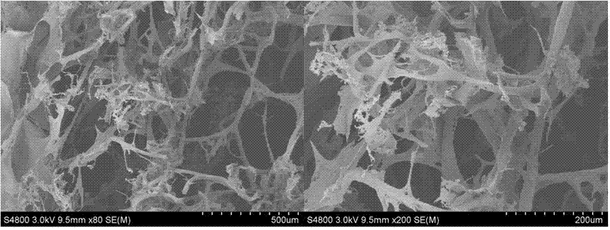
[0047] attached image 3 It is the scanning electron micrograph of the cross-section of the silk fibroin fiber support obtained above. It can be seen from the figure that the inside of the support is mainly composed of fibers, and the fibers hav...


PUM

PropertyMeasurementUnit
diameteraaaaaaaaaa
pore sizeaaaaaaaaaa
pore sizeaaaaaaaaaa

Abstract

The invention relates to a fibroin protein fiber scaffold and a preparation method thereof, after degumming of silk, the silk is soaked in an acid solution for dispersion to obtain a silk fiber dispersion solution; the silk fiber dispersion solution is injected into a mold for freezing to obtain a silk fiber frozen body; the silk fiber frozen body is immersed in an organic solvent to remove formic acid, and soaked and washed with deionized water to obtain a wet-state fibroin protein fiber scaffold; the-state fibroin protein fiber scaffold is coldly treated to obtain a frozen body, and the frozen body is frozen and dried to obtain the fibroin protein fiber scaffold. The internal structure of the fibroin protein fiber scaffold mainly comprises fiber, and the fibroin protein fiber scaffold has high porosity, high penetration rate and excellent mechanical properties, is very conducive to nutrient transport, cell migration and tissue growth, and is an ideal tissue engineering scaffold.

Description

technical field [0001] The invention relates to a silk fibroin fiber support and a preparation method thereof, which can be used in the fields of regenerative medicine such as repair of soft tissue and hard tissue, drug sustained release, and the like. Background technique [0002] There are millions of patients with organ or tissue damage and functional loss due to diseases and accidents every year. In the United States alone, more than 8 million operations are needed to treat such patients every year, and the economic cost is more than 400 billion US dollars. With the development of modern medicine and surgical techniques, tissue or organ transplantation to repair functional loss has been widely accepted, however, it faces a huge donor gap. The formation of tissues or organs in vivo or in vitro by means of regenerative medicine provides a new treatment option for the repair of damaged functions. Among them, the selection and construction of tissue engineering scaffold mat...

Claims


Application Information

Patent Timeline
Patent Type & Authority Applications(China)
IPC IPC(8): A61L27/22A61L27/50A61L27/56
CPCA61L27/227A61L27/50A61L27/56A61L2300/412
Inventor 张锋黄继伟左保齐
Owner 上海丝波敦生物科技有限公司
Why Eureka
Try Eureka