Magnetic fluorescent copolymer nano probe taking DNA (deoxyribonucleic acid) as template and application

A technology of nano-probes and copolymers, which is applied in the direction of fluorescence/phosphorescence, luminescent materials, and material analysis through optical means, which can solve the problems of high detection cost, expensive instruments and equipment, and low purity of circulating tumor cells. The effect of reducing the cost of detection and reducing the impact

Active Publication Date: 2018-07-06
SUZHOU UNIV
View PDF4 Cites 1 Cited by
  • Summary
  • Abstract
  • Description
  • Claims
  • Application Information

AI Technical Summary

Problems solved by technology

Although the CellSearch system has high capture efficiency and good repeatability, it also has some disadvantages: the equipment is expensive; because antibodies are used to capture...

Method used


Image

  • Magnetic fluorescent copolymer nano probe taking DNA (deoxyribonucleic acid) as template and application
  • Magnetic fluorescent copolymer nano probe taking DNA (deoxyribonucleic acid) as template and application
  • Magnetic fluorescent copolymer nano probe taking DNA (deoxyribonucleic acid) as template and application

Examples


Embodiment 1

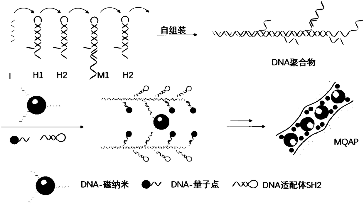
[0060] (1) Synthesis of DNA-quantum dots

[0061] In this embodiment, the C1DNA sequence used is as follows:

[0062] 5'-GGGGGGGGGGAAAAAA CGTCCGTGCTCAC -3', wherein base G has undergone thio modification.

[0063] The C2DNA sequence used is as follows:

[0064] 5'- TACGCGTCTAGGATC AAAAAAAAAAA-NH 2 -3'.

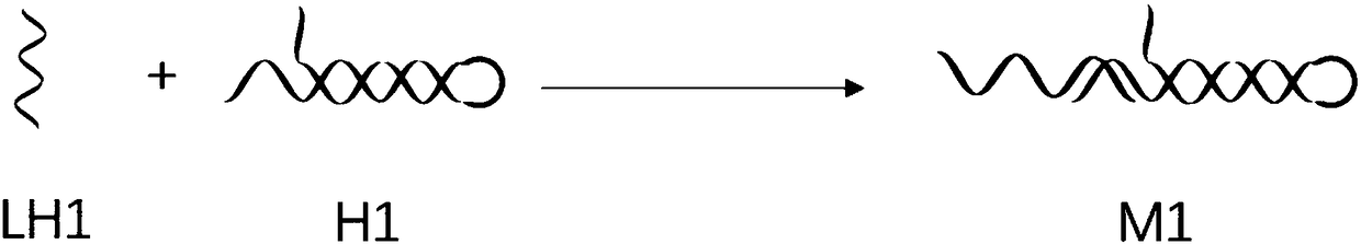
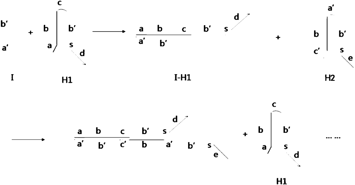
[0065] The H1 sequence used is as follows:

[0066] 5’-TTAACCCACGCCGAATCCTAGACTCAAAGTAGTCTAGGATTCGGCGTGAAAAA GTGAGCACGGACG -3', which is a hairpin structure, and the overhanging part is underlined.

[0067] The H2 sequence used was as follows:

[0068] 5'- GACGTGCAGGCTG TTTAAAGTCTAGGATTCGGCGTGGGTTAACACGCCGAATCCTAGACTACTTTG-3', which is a hairpin structure, and the overhanging part is underlined.

[0069] The promoter I sequence used is as follows:

[0070] 5'-AGTCTAGGATTCGGCGTGGGTTAA-3'.

[0071] The LH1 DNA sequence used was as follows:

[0072] 5'- GATCCTAGACGCGTA AAAAAAA CGTCCGTGCTCAC -3'.

[0073] The SH2 sequence used was as follows:

[0074] 5'- ...

Embodiment 2

[0085] Example 2 MQAP probes are used to specifically identify circulating tumor cells

[0086] (1) Culture of cell lines

[0087] The tumor cells to be detected, human acute lymphoblastic leukemia cells (CCRF-CEM) and the control cells, human B lymphocytoma cells (Ramos), were cultured. Both CCRF-CEM and Ramos cells are within 25cm 2 Cultured in the culture bottle of the culture medium, the culture medium is the RPMI 1640 culture fluid containing 1% double antibody (penicillin, streptomycin), 10% fetal bovine serum (FBS). The culture environment is 37°C, containing 5% CO 2 cell culture incubator.

[0088] (2) Specific recognition experiment of MQAP probe on CCRF-CEM cells

[0089] 200 μL of CCRF-CEM or Ramos cells (2×10 6 cells / mL) were first pre-stained with Calcein-AM (Calcein-AM, 10 μM) or 4,6-diamidino-2-phenylindole (DAPI, 10 μg / mL) in a 37°C water bath for 30 min, Add 500 μL of cell washing buffer to wash three times, and then react with 200 μL of the MQAP probe p...

Embodiment 3

[0091] Example 3 MQAP probes are used to separate and enrich CCRF-CEM cells

[0092] (1) According to the method of step (2) in Example 2, MQAP probes of different concentrations were interacted with CCRF-CEM cells, and then 100 μL (2×10 2 cells / mL) CCRF-CEM cells interacted with different concentrations of MQAP probes were added to a 24-well plate, a magnet was placed on the edge of the well plate for magnetic separation for 15 min, and then the captured tumor cells were counted by a fluorescence microscope. The cell separation device is attached Figure 7 as shown ( Figure 7 a is a schematic diagram of the separation device, Figure 7 b is the enlarged view of circle A in figure a, the arrow in the figure is the position where blood is added), and the fluorescence imaging characterization results captured by tumor cells are shown in the attached Figure 8 as shown, Figure 8 A is bright field, Figure 8 B is a quantum dot, and the arrow in the figure is a CCRF-CEM cell...


PUM


Abstract

The invention relates to a magnetic fluorescent copolymer nano probe taking DNA (deoxyribonucleic acid) as a template. The probe comprises a linear template DNA chain, wherein the template DNA chain is connected with magnetic nano particles with superparamagnetism, quantum dot nano particles with fluorescence and DNA or RNA (ribonucleic acid) aptamers capable of specifically identifying a tumor cell by identifying a DNA sequence; and the magnetic nano particles or the quantum dot nano particles and the DNA or RNA aptamers are alternately connected onto the template DNA chain. The invention further discloses an application of the magnetic fluorescent copolymer nano probe in a preparation for identifying a circulating tumor cell. The nano probe can amplify a magnetic signal and a fluorescentsignal, and has higher specific selection on tumor cells and lower non-specific absorption on normal blood cells.

Description

technical field [0001] The invention relates to the technical field of nano-biological materials, in particular to a magnetic fluorescent copolymer nano-probe with DNA as a template and its application. Background technique [0002] Circulating tumor cells (CTCs) play an important role in tumor metastasis, and 90% of tumor-induced deaths are due to tumor metastasis. Therefore, early diagnosis and treatment to prevent the spread of primary tumors play an important role in reducing mortality. Circulating tumor cells exist in the peripheral blood of patients in the early stages of cancer, so they can be used as important biological indicators for the diagnosis and treatment of cancer. However, the biggest difficulty at present is that the number of circulating tumor cells is extremely rare compared with normal cells in blood. Usually, the concentration of circulating tumor cells in the blood of cancer patients is only 1-3000 / ml, while the concentration of red blood cells is 10...

Claims


Application Information

Patent Timeline
IPC IPC(8): G01N21/64C09K11/06
CPCC09K11/06C09K2211/1466G01N21/6428G01N21/6486G01N2021/6439
Inventor 马楠李智
Owner SUZHOU UNIV
Why Eureka
Try Eureka