Double-signal non-enzyme signal amplification RNA (ribonucleic acid) nano-biosensor, preparation method and application of nano-biosensor
A biosensor and signal amplification technology, applied in the field of biosensors, can solve the problems of difficult detection and short miRNA length, and achieve the effects of high sensitivity, fast mass transfer rate, and small electrode size
- Summary
- Abstract
- Description
- Claims
- Application Information
AI Technical Summary
Problems solved by technology
Method used
Image
Examples
Embodiment 1
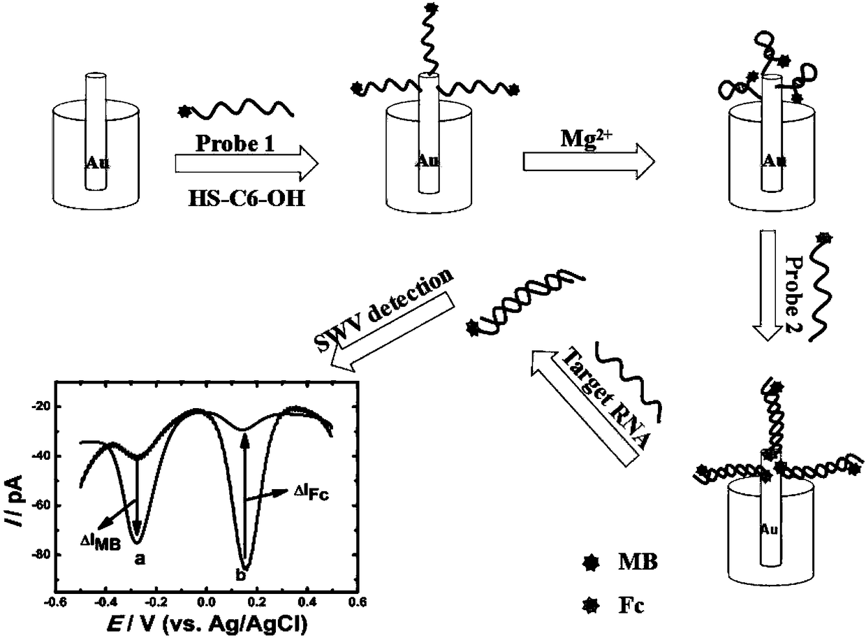
[0052] A method for preparing a dual-signal enzyme-free signal amplification RNA nano-biosensor, comprising the following steps:
[0053] 1) Dissolve 4mg of DTT in 100uL 0.1M, pH8.5 tris-HCl buffer solution to make a DTT buffer solution; mix the 1OD probe 1P-MB solution with the DTT buffer solution, and let stand at room temperature for 3 hours; then Add 200uL of 6% acetic acid solution and 50uL of 3M sodium acetate solution, add 1.6mL of 100% ethanol, mix well, centrifuge at 6000r / min, remove the supernatant, ventilate overnight at a constant temperature of 35°C, and dry to obtain Probe 1 after processing;
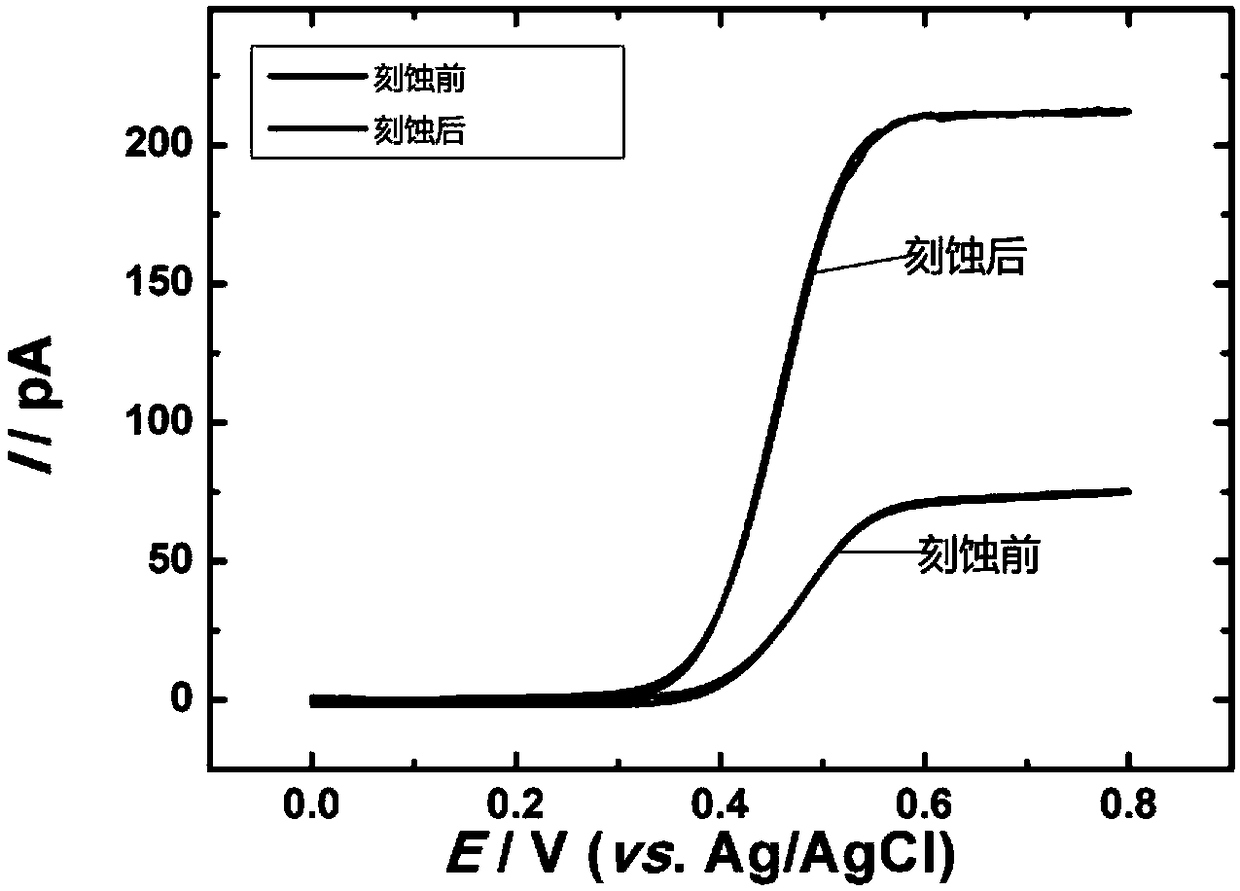
[0054] 2) Dissolve the probe 1 treated in step 1) with 0.01M PBS of pH 7.4, dilute it to 5uM, immerse the gold nanowire electrode with a diameter of 60nm prepared by the laser pulling method, and incubate at a constant temperature of 37°C for 12h Then, immerse the treated electrode in 100uL mercaptohexanol solution with a concentration of 10uM at room temperature, and se...
Embodiment 2
[0072] An application of a double-signal enzyme-free signal amplification RNA nano-biosensor to detect miRNA-16, specifically:
[0073] The Fc / MB / NEW electrode prepared in Example 1 was put into the prepared 200uL solution of target miRNA-16 with different concentrations. In order to enable better hybridization of the target miRNA, the selected conditions were hybridization at a constant temperature of 37° C. for 12 hours. The hybridized electrodes were mixed with 5mM MgCl 2 and 140mM NaCl in 0.01M PBS buffer solution, the SWV is as follows Figure 6A As shown, curve a is a blank control with a target concentration of 0. When the target miRNA concentration changes from 100fM to 100nM, it can be observed that the peak current of MB gradually increases, and the peak of Fc decreases accordingly. like Figure 6B Shown is the relationship between the current signal of methylene blue and ferrocene (after deducting the blank) and the concentration of target miRNA-16, and it is fou...
Embodiment 3
[0078] The prepared sensor was used for the trace detection of RNA in the environment of human blood samples. Add 50uL of serum to 10mL of 0.01M PBS buffer solution as the environment solution. Take 5mL of the environmental solution and add 5uL 5uM / L miRNA-16 standard solution, and incubate the Fc / MB / NEW electrode prepared in Example 1 with the sample for 12 hours. 2 and 140mM NaCl in 0.01M PBS buffer solution, and measured by square wave voltammetry. Afterwards, 5uL 5uM / L miRNA-16 standard solution was added to the solution in turn, and the electrochemical signal changes were recorded. The recovery rate was detected in the environment of human serum by the standard addition method, as shown in Table 1.
[0079] Table 1
[0080]
[0081] It can be seen from Table 1 that the recovery rate is maintained at about 96.0%-104.6%, indicating that the sensor prepared by the present invention can successfully achieve a sensitive response to miRNA-16 in a complex sample environmen...
PUM
 Abstract
Description
Claims
Application Information
 - R&D
- Intellectual Property
- Life Sciences
- Materials
- Tech Scout
- Unparalleled Data Quality
- Higher Quality Content
- 60% Fewer Hallucinations
Browse by: Latest US Patents, China's latest patents, Technical Efficacy Thesaurus, Application Domain, Technology Topic, Popular Technical Reports.
© 2025 PatSnap. All rights reserved.Legal|Privacy policy|Modern Slavery Act Transparency Statement|Sitemap|About US| Contact US: help@patsnap.com


